Online ISSN: 2187-2988 Print ISSN: 0911-1794
特定非営利活動法人日本小児循環器学会 Japanese Society of Pediatric Cardiology and Cardiac Surgery
Pediatric Cardiology and Cardiac Surgery 41(2): 84-88 (2025)
doi:10.9794/jspccs.41.84

症例報告Case Report

成人期に診断された動脈管動脈瘤に対してAmplatzer™ duct occluderを用いて経皮的閉鎖術を施行した1症例Transcatheter Closure of a Ductus Arteriosus Aneurysm Using an Amplatzer™ Duct Occluder in an Adult Patient

沖縄県立南部医療センター・こども医療センター 小児循環器内科Department of Pediatrics Cardiology, Nanbu Medical Center & Children’s Medical Center ◇ Okinawa, Japan

受付日:2024年12月25日Received: December 25, 2024
受理日:2025年1月27日Accepted: January 27, 2025
発行日:2025年5月31日Published: May 31, 2025
HTMLPDFEPUB3

動脈管動脈瘤(ductus arteriosus aneurysm: DAA)は遺残動脈管が嚢状または管状に拡張したもので,瘤破裂,血栓塞栓,感染などの致命的合併症を生じることがある.56歳男性において,閉鎖栓により経皮的に治療したDAA例を報告する.健康診断の胸部X線で左第1弓拡大を契機に,造影CT撮影からDAAと診断された.大動脈弓小弯側に入口部(径5.6 mm)をもつ最大径39 mmの嚢状動脈管瘤がみられ,肺動脈側に径2.2 mmの出口部がみられた.嚥下障害や嗄声など自覚症状はなかった.外科治療やstent graft挿入術の選択もあったが低侵襲治療として経皮的動脈管瘤閉鎖術を行った.大動脈側から挿入したguide wireを肺動脈側から挿入したsnare catheterで把持しwire loopを作成後,肺動脈側からdelivery sheathを挿入し,Amplatzer™ Duct Occluder 12 mm/10 mmにより閉鎖した.3カ月後の造影CTでは瘤内は血栓化されていた.経皮的閉鎖術は成人の巨大なDAAに対する有用な治療オプションの一つである.

A ductus arteriosus aneurysm (DAA) involves cystic or tubular dilatation of the persistent ductus arteriosus and can lead to life-threatening complications, including aneurysm rupture, thromboembolism, and infection. We report the case of a 56-year-old man who underwent percutaneous DAA closure. The patient was diagnosed with DAA following computed tomography (CT) scans after an abnormal finding on a routine chest radiography examination. CT imaging revealed an aneurysm with a maximum diameter of 39 mm, an aortic side diameter of 5.6 mm, and a pulmonary side diameter of 2.2 mm. The patient did not have any symptoms, such as dysphagia and hoarseness. Although surgical correction and stent graft implantation were considered, a minimally invasive percutaneous approach was chosen. A guidewire was introduced from the aortic side and retrieved with a snare catheter from the pulmonary artery (PA) side, creating a wire-loop. A delivery sheath was inserted from the PA side, and the aortic side of the DAA (measuring 5.6 mm) was percutaneously closed using a 12 mm/10 mm Amplatzer™ Duct Occluder (ADO). Follow-up CT conducted three months later confirmed near-complete thrombosis of the aneurysm. This case demonstrates that percutaneous closure of large DAAs in adults using devices such as the ADO is a safe, effective, and minimally invasive treatment option.

Key words: ductus arteriosus aneurysm; transcatheter closure; wire-loop technique; Amplatzer™ Duct Occluder

背景

動脈管動脈瘤(ductus arteriosus aneurysm: DAA)は動脈管が嚢状または管状に拡張を来す疾患である.動脈管の大動脈側が開存している動脈管憩室が進行性に拡大して瘤化すると考えられている1, 2).成人例は稀であるが,周囲構造への圧迫だけでなく,瘤破裂,血栓塞栓症,感染などの致死的合併症があるため早期の治療介入が必要である1, 2).治療法としては外科治療が施行されてきたが1, 3),近年ステントグラフトの報告もある4, 5).閉鎖栓を用いた経皮的閉鎖術の報告はほとんどなく,Amplatzer™ Duct Occluder(ADO)を用いた閉鎖報告はない.

56歳のDAA症例に対してADOを用いて経皮的閉鎖術を施行した.低侵襲かつ有用な治療オプションと思われるので報告する.

症例

症例

56歳,男性

現病歴

20歳頃に近院で動脈管開存と診断されたが,短絡量が少ないため治療適応にならなかった.その後通院を自己中断していた.54歳からII度高血圧の診断で降圧薬の内服を開始した.55歳時の健康診断で弓部大動脈瘤が疑われ,同院を受診した.造影CT像でDAA(瘤最大径39 mm,大動脈側入口部径5.6 mm,肺動脈側流出部径2.2 mm)と診断され,治療目的に当科に紹介された.瘤破裂の危険があるため,早期の介入治療を計画し,経皮的動脈管閉鎖術目的に入院した.

既往歴

緑内障,II度高血圧,高脂血症

家族歴

特記なし

生活歴

機会飲酒,喫煙なし

内服薬

アムロジピン10 mg/日,アテノロール25 mg/日,アトルバスタチン10 mg/日

来院時現症

身長160 cm,体重69.3 kg,体表面積1.73 m2,心拍数66/分,血圧127/80 mmHg,SpO2 97%(室内気),心音は整で心雑音は聴取せず,嗄声や嚥下障害は認められなかった.
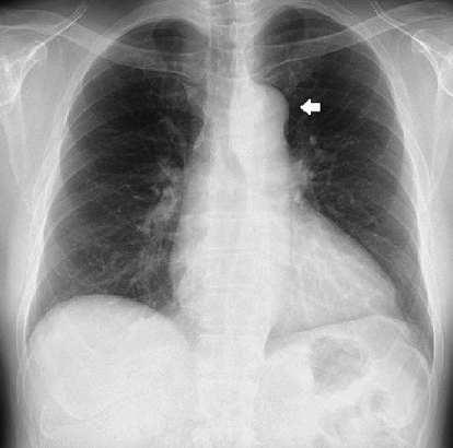
胸部X線

心胸郭比53%,左側大動脈弓陰影の突出を認めた(Fig. 1).

Pediatric Cardiology and Cardiac Surgery 41(2): 84-88 (2025)

Fig. 1 Chest X-ray showing the prominence of the left first cardiac arch due to ductus arteriosus aneurysm (DAA) (white arrow)

12誘導心電図

心拍数79/分,正常洞調律,QRS電気軸正常

心臓超音波検査

左室拡張末期径47 mm,左室駆出率70%,有意な弁逆流なし,腹大動脈パルスドプラ脈波 正常波形
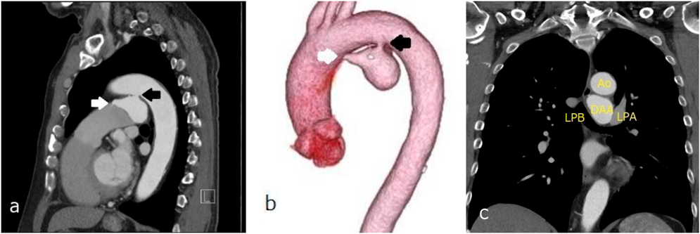
胸部造影CT

大動脈弓小彎側に入口部(径5.6 mm)をもつ最大径39 mの嚢状に拡大したDAAを認め,肺動脈側に出口部(2.2 mm)をもつ.大動脈解離なし.DAAは左主気管支と接触しているが圧排なし(Fig. 2).

Pediatric Cardiology and Cardiac Surgery 41(2): 84-88 (2025)

Fig. 2 Contrast-enhanced chest computed tomography revealing a DAA with a maximum diameter of 39 mm, an aortic side opening measuring 5.6 mm, and a pulmonary artery side opening measuring 2.2 mm

A fistula from the minor curve of the aortic arch to the aneurysm is presented (a, b: black arrows). Drainage from the aneurysm to the main pulmonary artery is indicated (a, b: white arrows). The left primary bronchus was in contact with DAA; however, it was not compressed (c). Ao, aorta; DAA, ductus arteriosus aneurysm; LPA, left pulmonary artery; LPB, left primary bronchus.

入院後経過

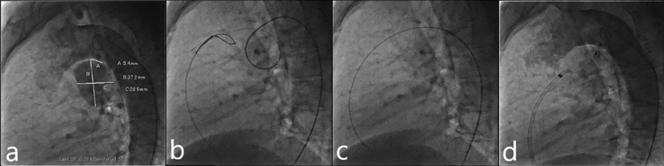
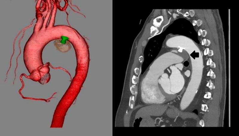
入院翌日に経皮的動脈管閉鎖術を施行した.局所麻酔後,右大腿静脈に6 Fr 11 cm sheath(Medikit Inc., Tokyo),右大腿動脈に5 Fr 11 cm sheath(Medikit Inc., Tokyo)を留置した.カテーテル検査の結果Qp/Qsは1.05,肺血管抵抗係数は0.57 Wood unit·m2.大動脈造影でDAAが造影され,瘤入口部径は5.4 mm,瘤最大径は28.9×37.2 mm,瘤流出部径は2.2 mmであった(Fig. 3a).5 Frジャドキンス型右冠動脈用(JR)カテーテル(Hanaco Medical, Saitama, Japan)を逆行性に進めて瘤入口部にかけて,そこから0.035̋Radifocus guidewire 260 cm(Terumo Inc., Tokyo)を瘤内へ進めて,1ループさせて主肺動脈まで進めた.続いて,順行性に進めて主肺動脈に留置した6 Fr wedge pressure catheter(Gadelius Medical, Kanagawa, Japan)から15 mm径の6 Fr Amplatz goose neck snare (Medtronic plc., Ireland)を進めて,Radifocus guidewire先端を把持して(Fig. 3b)右大腿静脈のsheath外まで引き出した.瘤内のループを伸ばして,大腿静脈–DAA–大腿動脈のwire loopを作成した(Fig. 3c).DAAの大動脈側入口部径が5.4 mmであったため,ADO 12 mm/10 mm(Abbott Structural Heart, Plymouth, MN, USA)を同部に留置した:右大腿静脈のsheathを抜去し,7 Fr Amplatzer™ TorqVue 180 (Abott, Pylmouth, MN, USA)を下行大動脈まで進めた.ADOのRetention skirtを展開して瘤入口部にかけて,デバイスボディを瘤内に展開した.留置後の大動脈造影では閉鎖栓突出による大動脈の狭窄はなく,遺残短絡はごく少量であった(Fig. 3d).特に合併症は認めず,治療翌日からアスピリン100 mgの内服を開始し,退院した.治療後の経過は良好で,術後1カ月の心臓超音波検査で遺残短絡は消失し,術後3カ月の胸部造影CT像で瘤内はほぼ血栓化していた(Fig. 4).

Pediatric Cardiology and Cardiac Surgery 41(2): 84-88 (2025)

Fig. 3 Percutaneous closure of a DAA

a: DAA size: Ao side, 5.6 mm; PA side, 2.2 mm; and maximum diameter, 37.2×28.9 mm. b: The distal end of the Radifocus guidewire is snared in the MPA. c: The Radifocus guidewire is withdrawn into the 6 Fr right femoral sheath to form a wire loop. d: After device deployment, aortic angiography shows no residual leakage from the DAA. DAA, ductus arteriosus aneurysm; MPA, main pulmonary artery.

Pediatric Cardiology and Cardiac Surgery 41(2): 84-88 (2025)

Fig. 4 Contrast-enhanced chest computed tomography revealing that the DAA is almost thrombosed (black arrow)

DAA, ductus arteriosus aneurysm.

考察

DAAは正期産児の8.8%に認められ,ほとんどが無治療で消退し,予後は良好である6).一方,成人期DAAは稀な疾患であり,動脈管の閉鎖過程において,肺動脈側の閉鎖に続く大動脈側の閉鎖遅延のため,動脈管が憩室として遺残し,これが次第に拡張して囊状動脈瘤を形成する疾患である3).本症例は肺動脈への交通が残存していたが,多くは肺動脈端が閉鎖している4).瘤化の原因としては,動脈管は大動脈や肺動脈よりも弾性線維に乏しく,中膜の粘液性変化もあるので7),体血圧を受けて徐々に拡大していくと考えられている2, 8).高齢,喫煙,男性,高血圧が動脈硬化性病変として影響している可能性もある1, 5)

成人期DAAは初めは無症状であるが,周囲構造を圧迫すると嗄声,咳嗽,呼吸苦,胸部痛,嚥下障害などが生じる.血痰,喀血は気管や食道への瘤穿破のサインである.合併症には破裂,解離,感染,塞栓症がある1–3, 5, 9).DAAの48%が瘤の合併症により死亡したという報告もあり1),DAAは放置すると致死率が高い疾患である.死亡例の瘤の最小サイズは4×3 cmと報告されている1).本症例は無症状であったが,破裂や塞栓症などの合併症のリスクを考慮して早期に治療介入した.

成人期DAAの治療に関しては,胸骨正中切開または左側開胸による開胸手術が一般的であり,閉鎖栓を用いた経皮的閉鎖術の報告はほとんどない.開胸手術では人工心肺を用いた瘤切除,交通孔の直接閉鎖もしくはパッチ閉鎖,人工血管置換が施行されるているが1, 3, 5),皮膚切開の範囲が大きく,血胸,乳び胸や反回神経損傷などのリスクがある.また,動脈硬化による石灰化を伴っている場合は血管壁の脆弱性から直接閉鎖は難易度が高くなる.近年,大動脈ステントグラフト内挿術(Thoracic endovascular aneurysm repair: TEVAR)も施行されているが4, 5),22 Fr程度の大口径sheathを用いるため通常カットダウン法によるアプローチが必要となる.また,重大な合併症に脳梗塞や脊髄虚血による対麻痺が挙げられる.

一方,閉鎖栓を用いた閉鎖術は大腿動脈または大腿静脈から6~7 Fr sheathを用いて施行可能であり,出血量は少なく,入院期間も短い低侵襲な治療法である.18×14 mmとそれほど大きくはないDAAの中にAmplatzer™ vascular plug II (AVPII)を留置した報告はあるが10),ADOを用いた閉鎖報告は本報告が初と思われる.本症例のDAAは最大径28.9×37.2 mmと大きいが,大動脈からの流入部径は5.4 mmとそれほど太くはなく,また肺動脈への流出部が残存していたので,大腿動脈–DAA–大腿静脈ワイヤーループが作成可能であり,順行性に瘤入口部をADOを用いて閉鎖できた.肺動脈への流出部を閉鎖した場合は,動脈瘤内に血液が流入する圧負荷が持続して瘤がさらに拡大する危険があるので,大動脈側を閉鎖する必要がある.また,瘤流出部が閉塞している場合はADOを留置できないので,AVPIIや,径が大きい場合は心房中隔欠損閉鎖用の閉鎖栓を逆行性に瘤流入部に留置する手段が考えられる.また,瘤内をコイルで閉鎖する方法もありうるが,大きな瘤を閉鎖する場合は多くのコイルが必要になり,また手技時間も長くなると予想されるので,閉鎖栓を用いた閉鎖が望ましい.

閉鎖栓留置後の抗凝固・抗血小板療法に関するガイドラインはないが,瘤入口部をADOなどの閉鎖栓で閉鎖した場合,大動脈側は通常の動脈管を閉鎖した場合と同様なので薬剤は不要と思われる.しかし,瘤内で生じた血栓が肺動脈へ流れて肺塞栓を来す可能性があるので,血栓化・内膜化が進むまでの間は抗血小板薬の使用が望ましいと思われる.本症例では1年間アスピリンを使用した.

結語

DAAは成人期に診断されることは稀であるが,破裂や塞栓症の合併が多いことから早期の治療介入が望ましい.本症例のようにDAAの形態からADOを用いた閉鎖が可能なものが存在する.DAAに対するADOなどの閉鎖栓を用いた経皮的閉鎖術は低侵襲かつ短期間の入院で治療可能であり,有用な治療選択肢の1つである.

倫理的配慮・説明と同意

論文投稿について本人に説明し,同意を得ている.

利益相反

本論文について開示すべき利益相反(COI)はない.

著者の役割

担当医,論文著者:西岡真樹子

論文指導:北野正尚,佐藤誠一

診療協力者:島袋篤哉,吉野佳佑

引用文献References

1) Mitchell RS, Seifert FC, Miller DC, et al: Aneurysm of the diverticulum of the ductus arteriosus in the adult. Successful surgical treatment in five patients and review of the literature. J Thorac Cardiovasc Surg 1983; 86: 400–408

2) Cruickshank B, Marquis RM: Spontaneous aneurysm of the ductus arteriosus: A review and report of the tenth adult case. Am J Med 1958; 25: 140–149

3) Ueno Y, Shinozaki T, Shimamoto M, et al: Aneurysm of the diverticulum of the ductus arteriosus in the adult. Nihon Kyobu Geka Gakkai Zasshi 1990; 38: 1356–1361

4) Saito N, Kimura T, Toma M, et al: Successful endovascular repair of an aneurysm of the ductus diverticulum with a branched stent graft: Case report and review of literature. J Vasc Surg 2004; 40: 1228–1233

5) Yukimoto M, Okuma T, Sohgawa E, et al: Incidentally identified ductus arteriosus aneurysm in eight adults: A case series. BJR Case Rep 2021; 7: 20200097

6) Jan SL, Hwang B, Fu YC, et al: Isolated neonatal ductus arteriosus aneurysm. J Am Coll Cardiol 2002; 39: 342–347

7) Ohtsuka S, Kakihana M, Ishikawa T, et al: Aneurysm of patent ductus arteriosus in an adult case: Findings of cardiac catheterization, angiography, and pathology. Clin Cardiol 1987; 10: 537–540

8) Lund JT, Jensen MB, Hjelms E: Aneurysm of the ductus arteriosus: A review of the literature and the surgical implications. Eur J Cardiothorac Surg 1991; 5: 566–570

9) Tsujimoto S, Hirose K, Ohyagi A: A ruptured large aneurysm of the ductus arteriosus. Heart 1987; 57: 289–291

10) van der Linde D, Witsenburg M, van de Laar I, et al: Saccular aneurysm within a persistent ductus arteriosus. Lancet 2012; 379: e33

This page was created on 2025-04-25T13:09:34.655+09:00
This page was last modified on 2025-07-01T14:00:05.000+09:00


このサイトは(株)国際文献社によって運用されています。