動脈管の収縮形態:ラット動脈管断面像天然色図譜Natural Color Cross-Sectional Morphology of Constricted Ductus Arteriosus in Rat
東京女子医科大学循環器小児科Department of Pediatric Cardiology, Tokyo Women’s Medical University ◇ Tokyo, Japan
発行日:2018年9月1日Published: September 1, 2018
生体内動脈管の収縮を研究するため,ラット胎仔と新生仔を全身急速凍結法で固定し凍結ミクロトームで胸部を矢状面,前額面,横断面,または四腔断面で切り,0.5 mmごとに断面カラー写真を連続撮影した.胎仔の矢状面で胎生期主要血流路である右室漏斗部–主肺動脈–動脈管–下降大動脈が一断面に同じ太さで明示された.新生仔の矢状面で生後30分の動脈管の管状収縮が明示され,出生後90分で速やかに管状の狭窄を経て閉鎖した.動脈管索は生後3日で著明な短縮を生じた.胎生期動脈管の薬剤性収縮は新生仔の動脈管収縮と異なる形態を示した.即ちインドメサシンを親ラットに経口投与すると4時間後の中央部に強い砂時計型収縮,8~24時間後の限局性の大動脈側の膜状収縮が明示された.この特徴ある胎生期動脈管収縮は全て薬剤による収縮,即ちインドメサシンなど鎮痛解熱剤,ベタメサゾンなどステロイドホルモン,グリベンクラミドなどのスルホニル尿素薬によるラット胎仔動脈管収縮で観察された.胎仔動脈管収縮24時間持続後の四腔断面には右室の内腔狭小化と壁肥大,左室の拡張と肥大,心嚢液貯留が明示された.
In-situ cross-sectional morphology of the fetal and neonatal rat ductus arteriosus (DA) is studied by rapid whole-body freezing, cutting on a freezing microtome, and photographing the cross-section serially every 0.5 mm with a stereoscopic microscope (Wild M400). Thoracic sagittal sections reveal the right ventricular (RV) infundibulum, main pulmonary artery, DA, and descending aorta in one plane. This forms the major circulatory route in the fetus. Serial frontal and sagittal sections of the neonatal thorax reveal rapid tubular constriction at 1 hour and complete closure at 2 hours after birth. Fetal DA shows different patterns of pharmacological constriction. Fetal DA constriction by indomethacin is initially tubular at 1 and 2 hours after orogastric administration to the mother rat but becomes sandglass shaped and eventually becomes distal, short, tubular, or membranous at 8 and 24 hours after administration. These patterns of constriction are observed after pharmacological agents are administered transplacentally or directly to the fetus; this includes DA constriction after administration of cyclooxygenase inhibitors such as aspirin, indomethacin, and ibuprofen, constriction after administration of glucocorticoid hormones such as betamethasone, and constriction after administration of sulfonylureas such as glibenclamide and glimepiride. Persistent fetal DA constriction after administration of indomethacin for more than 24 hours induces RV concentric hypertrophy with diminished cavity and left ventricular dilatation with increased ventricular muscle mass in 4-chamber view sections.
Key words: fetus; ductus arteriosus; ductus constriction; ductus closure; indomethacin
© 2018 特定非営利活動法人日本小児循環器学会© 2018 Japanese Society of Pediatric Cardiology and Cardiac Surgery
2017年のノーベル化学賞1)はクライオ電子顕微鏡の開発に与えられた.生体構造の観察と計測に凍結は最良の固定法である.ラット胎仔と新生仔の全身急速凍結法による凍結標本を凍結ミクロトームで切り,その断面の心臓大血管像から種々の計測を行う方法は50年前に動脈管の研究用にKarolinska研究所で開発され2),40年前から私も用いてきた3–10).この方法は胎仔出生前後の心臓と大血管の生理的形態変化の研究11),先天性心疾患の形態研究12–17)と心不全の評価18)にも用いられ,臨床上の胎児エコーによる心臓断面像よりも鮮明な天然色心臓断面を提示できる.これらの天然色の断面写真はヒトでは観察できない細部迄の解剖が記録されており,ヒト胎児のモデルとしても興味がある.従来は印刷費用の制約からこれらのカラー写真の論文発表は一部分にとどまったが,本誌の電子書籍化でその制約がなくなったので,ここに動脈管を中心に図譜にして発表したい.なお胎生期先天性心疾患のカラー写真は別にまとめた12).また卵円孔と静脈管については別の図譜にまとめる予定である.それぞれの実験の計測データーは紙面の関係で省略するので,原著3–11, 18–21)を参照されたい.
ラット満期胎仔の全身急速凍結法による実験3–21)を次のように行った.ラットの妊娠期間は21.5日なので,妊娠21日目に親ラットを頸椎脱臼法で安楽死せしめ,ただちに帝王切開で取り出した胎仔を胎盤つきのままドライアイス–アセトン(−76°C)に投入して瞬時に凍結した.凍結した胸部を凍結ミクロトームで横断面transverse section,前後面sagittal,前額面frontalまたは四腔断面4 chamber viewで切り,0.1ないし0.5 mmごとに実体顕微鏡(Wild M400)とカラーフィルム(Reale,富士フィルム)で撮影した.1個体の胸部から約20断面を撮影した.10年にわたる実験の途中で光源とフィルターを変更したので全体の色調に差が生じた.出生直後の新生仔は妊娠21日目に親ラットの頸椎脱臼と帝王切開でとりだした新生仔を34°Cの保温器で育て,1定時間後の実験に用いた.生後1~4日後のラットは予定日に自然出産後に親の保育授乳中のラットを用いた.インドメサシンによる動脈管収縮は妊娠21日目に親ラットにインドメサシン10 mg/kgを胃内注入し,一定時間後に胎仔を全身急速凍結法で固定した.
凍結ミクロトーム上の切断面は次のように選択した.動脈管の研究では矢状面と前額面を用いた.Figs. 1, 2, 5, 6の動脈管矢状面はFig. 1Cに示すように矢状面より10~15度左寄りである.Fig. 7の動脈管横断面は脊柱に平行な前額面である.
Abbreviations in Figures 1–10. A: anterior, Ao: aorta, AoV: Aortic valve, Ca: caudal, Cr: cranial, DA: ductus arteriosus, DAo: descending aorta, DV: ductus venosus, E: esophagus, FO: foramen ovale, HV: hepatic vein, I: inferior (caudal), IAS: interatrial septum, LA: left atrium, LB: left bronchus, LCA: left coronary artery, LPA: left pulmonary artery, LSVC: left superior vena cava, LV: left ventricle, MPA: main pulmonary artery, P: posterior, PFO: patent foramen ovale, PoV: portal vein, PV: pulmonary vein, PVa: pulmonary valve, RA: right atrium, RB: right bronchus, RPA: right pulmonary artery, RV: right ventricle, RVI: right ventricular infundibulum, S: superior (cranial), SVC: superior vena cava, T: trachea, US: umbilical sinus, UV: umbilical vein
ラットの解剖学書22)によるとラットとヒトでは心臓と胸部大血管の解剖は大体共通しているが,次の種差がある.冠状静脈洞に還流する左上大静脈はヒトでは疾患として生じるがラットなどげっ歯類では正常である.左右の肺静脈はヒトでは左右2本ずつ左房に入るが,ラットでは左房後方で合流して共通肺静脈になり,左房に接続する.
心臓血管断面写真.(カッコ内に類似写真を白黒写真で発表した論文の文献番号を示した)
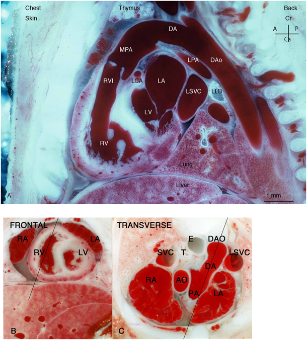
Fig. 1は胎生期の主要血流路23)である右室流出路–主肺動脈–動脈管–下降大動脈の矢状面断面の写真である.胎児エコー上でのductal archに相当する.ほぼ同じ太さの主肺動脈と動脈管と下行大動脈11),および細い左肺動脈11)を示す(初出).
Fig. 2は胎仔の矢状面の連続断面である.Fig. 1とわずかに異なる角度で切れている.動脈管径の計測にはFig. 7に示す前額面断面を用いた(初出).
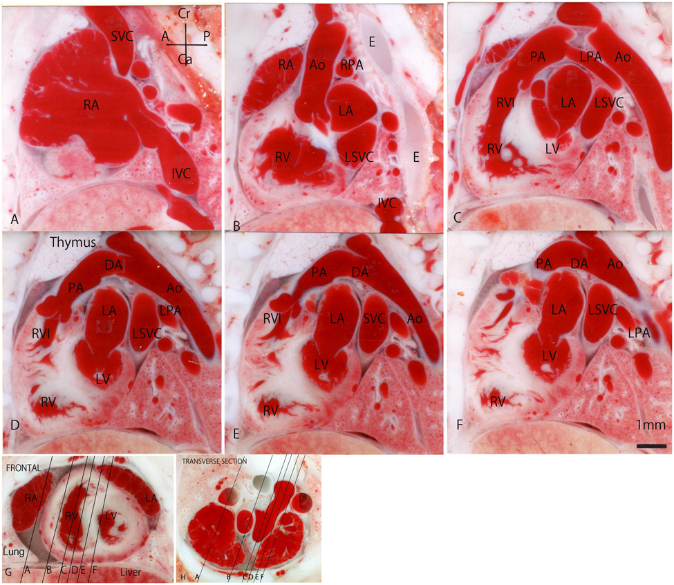
Fig. 3は生後1日新生仔心臓後部と肝臓の前額面,Fig. 4は生後4日の新生仔である.動脈管は閉じて索状になり,卵円孔も閉じ,左右肺動脈の急速な生後の拡大11)がある.
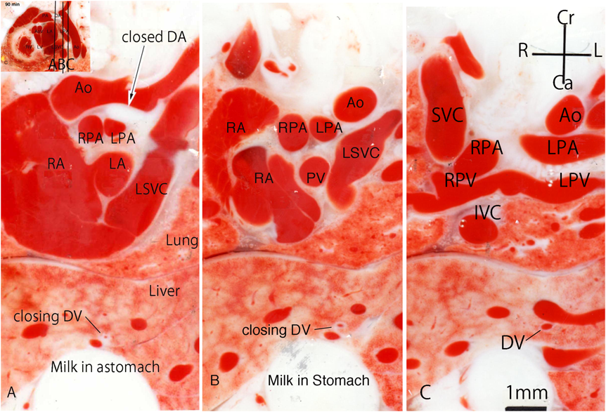

Fig. 5はラット新生仔の動脈管矢状面である.生後10分で軽度管状に収縮し,60分で高度に収縮し,90分で完全に閉鎖する11).
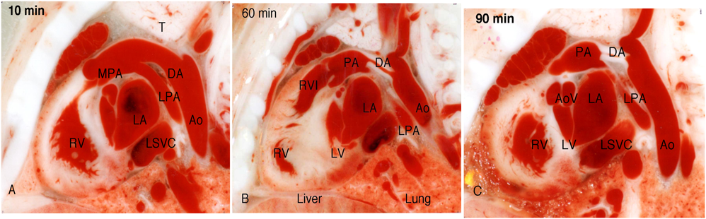
Fig. 6はラット胎仔のインドメサシン(10 mg/kg,親ラット胃内注入)による収縮を示す.4時間後(A)には砂時計型の収縮があり,24時間後には大動脈端に膜型(B),または短い管型(C)の狭窄が残った.Cには右室肥大と内腔狭小化,心嚢液の貯留がある4–10, 18, 19).ヒトでもアセチルサリチル酸による満期胎児の動脈管の大動脈接続部の膜性閉鎖例の報告24)がある.
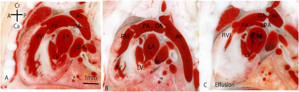
A: At 4 hours. B, C: at 24 hours. C shows concentric hypertrophy of the right ventricle and effusion
Fig. 7はラット胎仔のインドメサシン(10 mg/kg,親ラット胃内注入)による収縮24時間後の前額面断面である.動脈管横断面で顕微鏡とマイクロメーターで内径を測定し,通常内径最小値で動脈管収縮を表す3–10).

Fig. 8は正常ラット胎仔の横断面である(初出).
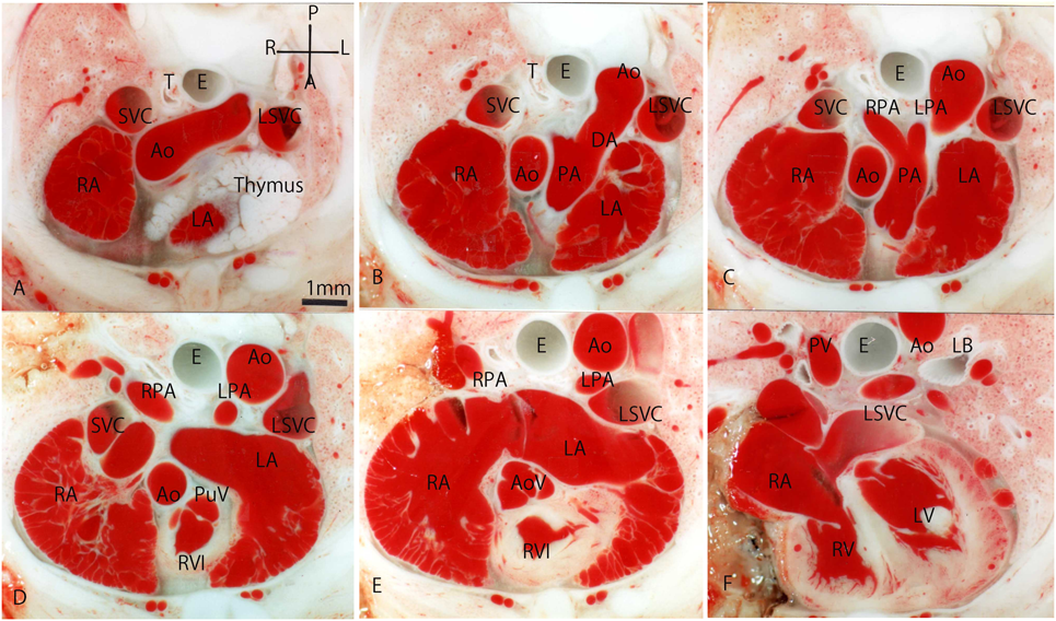
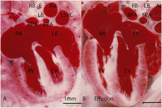
Fig. 9は胎仔の横断面で,親ラットにインドメサシン1 mg/kg,べタメサゾン(1 mg/kg)を投与して2時間後の胎仔動脈管収縮を示す.動脈管収縮以外に目立つ変化はない20).

Fig.10Aは対照,Bはインドメサシン10 mg/kgを親ラットに投与して24時間後のラット胎仔心臓の四腔断面である.Bは対照と比べ,右室の内腔縮小と壁肥大,左室の内腔拡大を示す.この実験は右室低形成を伴う純型肺動脈弁閉鎖症の発生モデルになる4).
動脈管の臨床では,胎生期早期閉鎖,未熟児の開存など多くの研究テーマが残っている.この動脈管図譜を通じて動脈管収縮の超音波診断が更に進むことを期待している.
本稿において利益相反に関して記載すべき事項はありません.
1) 朝日新聞:ノーベル化学賞に欧米3氏:高精密な電子顕微鏡開発.朝日新聞デジタル(2017年10月4日)
2) Hörnblad PY, Larsson KS: Studies on closure of the ductus arteriosus. I. Whole-body freezing as improvement of fixation procedures. Cardiologia 1967; 51: 231–241
3) Momma K, Toyoshima K, Takeuchi D, et al: In vivo constriction of the fetal and neonatal ductus arteriosus by a prostanoid EP4-receptor antagonist in rat. Pediatr Res 2005; 58: 971–975
4) Momma K, Takao A: Right ventricular concentric hypertrophy and left ventricular dilatation by ductal constriction in fetal rat. Circ Res 1989; 64: 1137–1146
5) Momma K, Takao A: Transplacental cardiovascular effects of four popular analgesics in rats. Am J Obstet Gynecol 1990; 162: 1304–1310
6) Momma K: Fetal and neonatal ductus arteriosus, in Cutis-Prior P (ed): The Eicosanoids. The John Wiley & Sons, 2004, pp 569–581
7) Toyoshima K, Momma K, Nakanishi T: In vitro dilatation of the ducts arteriosus induced by furosemide in the rat. Pediatr Res 2010; 67: 173–176
8) Momma K, Toyono M: The role of nitric oxide in dilating the fetal ductus arteriosus in rats. Pediatr Res 1999; 46: 311–315
9) Momma K, Toyoshima K, Takeuchi D, et al: In vivo constriction of fetal and neonatal ductus arteriosus by a prostanoid EP4-receptor antagonist in rats. Pediatr Res 2005; 58: 971–975
10) 門間和夫:動脈管薬の実験40年.日小児循環器会誌2016; 32: 261–269
11) Momma K, Takao A, Ito R, et al: In situ morphology of the heart and great vessels in fetal and newborn rats. Pediatr Res 1987; 22: 573–580
12) 門間和夫:ラット胎仔先天性心疾患の断面像—胎児心エコーのための染色体22q11.2欠失症候群モデル動物図譜として—.日小児循環器会誌2018; 34: 55–62
13) Momma K, Ando M, Takao A: Fetal cardiac morphology of tetralogy of Fallot with absent pulmonary valve in the rat. Circulation 1990; 82: 1343–1351
14) Momma K, Ando M, Takao A, et al: Fetal cardiovascular morphology of truncus arteriosus with or without truncal valve insufficiency in the rat. Circulation 1991; 83: 2094–2100
15) Momma K, Ando M, Takao A, et al: Fetal cardiovascular cross-sectional morphology of tetralogy of Fallot in rats. Fetal Diagn Ther 1990; 5: 196–204
16) Momma K, Ando M: Morphology of atrioventricular septal defect and fetal hydrops in the fetal rat. Cardiol Young 1993; 3: 13–19
17) Momma K, Ando M: Fetal cardiovascular morphology of interrupted aortic arch type B in rats. Fetal Diagn Ther 1994; 9: 44–52
18) 門間和夫,森 善樹,山村英司:胎生期心不全の心臓断面形態.新生児誌1995; 27: 626–632
19) Momma K, Konishi T, Hagiwara H: Characteristic morphology of the constricted fetal ductus arteriosus following maternal administration of indomethacin. Pediatr Res 1985; 19: 493–500
20) Momma K, Takao A: Increased constriction of the ductus arteriosus with combined administration of indomethacin and betamethasone in fetal rats. Pediatr Res 1989; 25: 69–75
21) Momma K, Ando S: In situ morphology of fetal aortic isthmus following ductal constriction in rats. Fetal Diagn Ther 1994; 9: 53–61
22) Hebel R, Stromberg MW: Anatomy and Embryology of the Laboratory rat. Wörthsee, BioMed Verlag, 1986, pp 112–113
23) Rudolph AM: Congenital Diseases of The Heart: Clinical-Physiological Considerations, Third Ed., Chichester, UK, Wiley-Blackwell, 2009, pp 1–24
24) Arcilla RA, Thilenius OG, Ranniger K: Congestive heart failure from suspected ductus closure in utero. J Pediatr 1969; 75: 74–78


This page was created on 2018-07-20T17:05:03.574+09:00
This page was last modified on 2018-09-11T14:52:54.354+09:00
このサイトは(株)国際文献社によって運用されています。