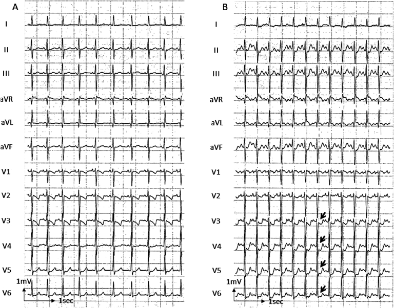
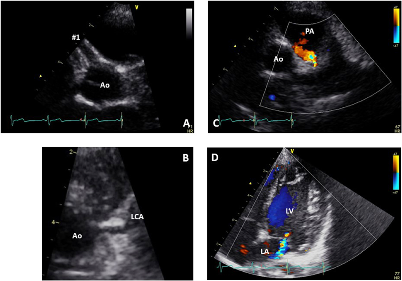
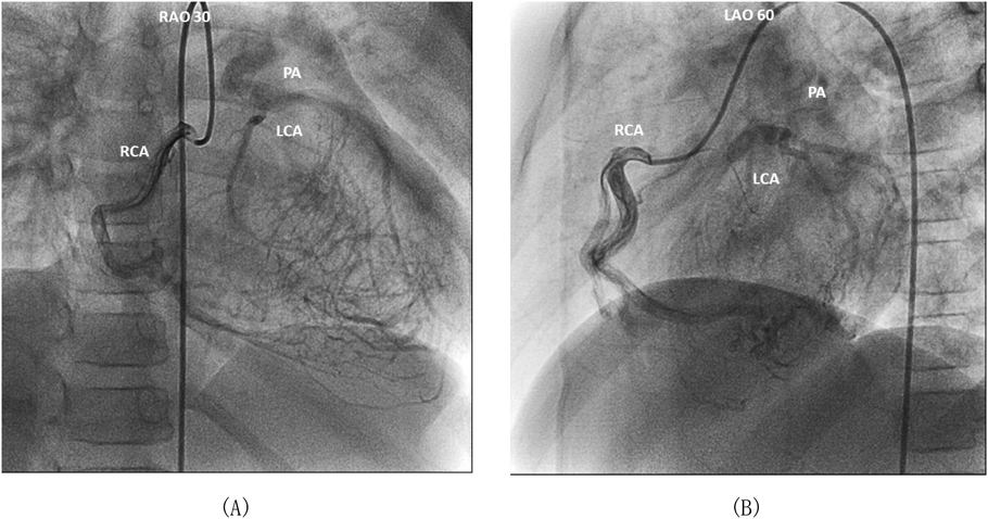
Online ISSN: 2187-2988 Print ISSN: 0911-1794
特定非営利活動法人日本小児循環器学会 Japanese Society of Pediatric Cardiology and Cardiac Surgery
Pediatric Cardiology and Cardiac Surgery 35(1): 38-42 (2019)
doi:10.9794/jspccs.35.38

症例報告Case Report

急性期川崎病診療のピットフォール左冠動脈肺動脈起始症Pitfall in Acute Care of Kawasaki Disease: Anomalous Origin of the Left Coronary Artery from the Pulmonary Artery

久留米大学医学部小児科学教室Department of Pediatrics and Child Health, Kurume University School of Medicine ◇ Fukuoka, Japan

受付日:2018年4月16日Received: April 16, 2018
受理日:2018年11月8日Accepted: November 8, 2018
発行日:2019年3月1日Published: March 1, 2019
HTMLPDFEPUB3

川崎病は日常診療で遭遇する機会が多く,合併症として冠動脈拡大・瘤の診断は重要である.一方,冠動脈拡大・瘤を示す疾患には,左冠動脈肺動脈起始症,冠動脈瘻,左冠動脈閉鎖症などの先天性冠動脈異常もある.症例は6歳女児で,歳時に川崎病に罹患し,前医に入院した.免疫グロブリン大量療法を施行され,速やかに解熱したが,初回心エコー図検査時より右冠動脈拡大があり,縮小することなく残存した.川崎病性右冠動脈拡大,軽症僧帽弁閉鎖不全,動脈管開存症疑いとして前医で外来管理されていた.5歳時より全力疾走時に胸痛を訴えるようになった.運動負荷心電図検査を施行したところ,自覚症状はなかったが,V3–6誘導で0.1–0.2 mVの下降型ST低下が認められ,精査加療目的で当院紹介された.当院で冠動脈造影CT検査を施行し,左冠動脈肺動脈起始症と確定診断した.術前の精査では左室前側壁領域の軽度壁運動低下を認め,左冠動脈移植手術を行った.今回,川崎病性右冠動脈拡大と診断され,無治療経過観察されていた左冠動脈肺動脈起始症の1小児例を経験した.急性期川崎病診療のピットフォールとして,注意喚起すべきと考えたので報告する.

Kawasaki disease (KD) is the most prevalent acquired vasculitis in children; coronary artery dilation and aneurysms are the most important complications of this disease. Alternatively, coronary artery dilation can be observed in rare congenital coronary artery anomalies, such as anomalous origin of the left coronary artery from the pulmonary artery (ALCAPA). At the age of 3, the patient suffered from typical KD, which was successfully treated with immunoglobulin infusion; however, right coronary dilation was noted at 4 days of illness. She was followed-up for the persistent right coronary artery dilation. At the age of 6, she complained of chest pain, and exercise stress test revealed ST segment depression in leads V3–V6, which resulted in the tentative diagnosis of coronary artery fistula. She was referred to our hospital for further evaluation and treatment. X-ray coronary computed tomography lead to the diagnosis of ALCAPA and stress myocardial scintigraphy confirmed ischemia of the anterolateral wall of the left ventricle; therefore, she underwent successful direct reimplantation of the anomalous left coronary artery to the aortic root. Here, we report the case of a patient with ALCAPA who was misdiagnosed as having coronary artery dilation associated with KD, and the patient was followed-up without appropriate treatment. We found that misdiagnosis of congenital anomalies can be a drawback of echocardiographic evaluation of coronary arteries in patients with acute KD, and clinicians must be able to properly distinguish between both the plausible causes of dilation.

Key words: coronary artery aneurysm; anomalous origin of the left coronary artery from the pulmonary artery; Kawasaki disease; echocardiography

はじめに

川崎病は,日本での年間患者数は約1万5千人と小児科日常診療で遭遇する機会が多い.罹患者の約3%に冠動脈拡大・瘤が残存し1),ガイドライン2)に則って診断とフォローアップが行われている.小児科医にとって,川崎病は,冠動脈拡大・瘤を有する小児患者を診たときに,もっとも考慮する疾患である.

一方,冠動脈拡大・瘤を示す疾患として,先天性冠動脈異常がある.先天性冠動脈異常は①冠動脈起始部位と走行形態の異常,②冠動脈開口部の狭窄・閉鎖,③冠動脈血管自体の異常,④冠動脈終末端の異常の大きく4つに分類される.冠動脈の走行には個人差があり,先天性冠動脈異常をどのように定義するかは報告によって異なるが,頻度は約1%とされ稀ではない3).このなかでも左冠動脈肺動脈起始症(anomalous origin of the left coronary artery from the pulmonary artery:ALCAPA)などの一部の異常は,心筋梗塞,不整脈,心不全などを来たして生命予後に影響する.したがって冠動脈拡大・瘤を見たときには,先天性冠動脈異常を念頭に置く必要がある4, 5)

今回,我々は川崎病急性期診療中に川崎病性右冠動脈拡大と診断され経過観察されていたALCAPAの1小児例を経験した.冠動脈拡大・瘤を有する小児患者であっても川崎病によるものと早計に診断せず,ALCAPAなどの先天性冠動脈異常を念頭に置いて心エコー図検査・心電図検査を行うことを啓蒙する必要があると考え報告する.

症例

症例

6歳女児

紹介理由

冠動脈病変の精査加療目的

現病歴

3歳1か月,川崎病に罹患(主要症状:6/6項目),第4病日(初診時)の心エコー図検査で右冠動脈拡大(#1径2.7 mm),軽度僧帽弁閉鎖不全,動脈管開存によると思われる肺動脈への流入血流を指摘された.前医で免疫グロブリン大量療法施行され,8病日に解熱した.以後,特に症状なく,前医外来定期管理されていた.

しかし,5歳時より全力疾走時に胸痛を訴えるようになった.胸痛の部位は胸部正中で,安静にすると数分で自然軽快するようなものであった.また,右冠動脈拡大は経年的に進行していった(4歳時 #1径3.0 mm,5歳時 #1径3.5 mm).6歳4か月時,胸痛と右冠動脈拡大,肺動脈への流入血流から,冠動脈瘻を疑われ,前医でトレッドミル運動負荷心電図検査(Fig. 1)を施行された.胸痛は訴えなかったが,V3–6で0.1–0.2 mVの下降型ST低下が認められたため,1か月後,冠動脈病変の精査加療目的に当院紹介された.

Pediatric Cardiology and Cardiac Surgery 35(1): 38-42 (2019)

Fig. 1 Exercise stress electrocardiograms

(A) The 12-lead electrocardiogram at rest. (B) The 12-lead electrocardiogram during exercise stress test at a workload of 8.4 METS. Exercise induced ST segment depression in leads V3–V6 (arrows).

既往歴

特記事項なし

家族歴

父方祖父に拡張型心筋症

入院時現症

身長:107.7 cm(-1.7SD),体重:18.2 kg(-0.9SD),心拍数:83 bpm,血圧:94/65 mmHg,呼吸数:20回/分,心音整,心雑音なし,奔馬調律なし,腹部・四肢に特記すべき所見なし.

血液検査

NT pro-BNP 174.6 pg/mL, CK 85U/L.その他,特記事項なし.

胸部X線検査

心胸郭比0.49,肺うっ血なし.

12誘導心電図

洞調律,97 bpm,左軸偏位,PQ時間0.11秒,QRS幅0.08秒,異常Q波なし,I・aVL・V5・V6誘導で0.1 mV以内の水平型ST低下,aVL誘導で陰性T波.

心エコー図検査(Fig. 2)

Pediatric Cardiology and Cardiac Surgery 35(1): 38-42 (2019)

Fig. 2 Echocardiographic findings of the patient at the first visit to our institution

(A) Parasternal short axis view of the aortic root shows right coronary artery dilation with a diameter of 4.6 mm at #1. (B) In this view, the left coronary artery appears to be normally connected with the aorta. (C) Color Doppler echocardiography of tilted parasternal short axis view shows abnormal flow into the main pulmonary artery. (D) Color Doppler echocardiography at the apical four-chamber view shows mild mitral regurgitation. Ao, aorta; LA, left atrium; LCA, left coronary artery; LV, left ventricle; PA, pulmonary artery.

左室拡張末期径35.1 mm(+0.6SD),左室壁運動異常なし,左室駆出率75%.軽度の僧帽弁閉鎖不全を認めたが,僧帽弁逸脱は認めなかった.主肺動脈への拡張期流入血流を認めたが,大動脈との連続性がないため動脈管血流とは考えられず,冠動脈瘻等の異常血流の可能性を考えた.

冠動脈は#1 径4.6 mm(+6.73SD),#2 径3.3 mm,#3 径1.6 mm,#5 径2.3 mm(+0.22SD)と,右冠動脈は全体的に拡大しており,川崎病急性期より増大していた.左冠動脈は,大動脈基部より正常に起始しているかのように描出されたが(Fig. 2B),はっきりしなかった.

経過

冠動脈造影CTで左冠動脈が肺動脈から起始する所見が得られ,ALCAPAと確定診断した.心臓カテーテル検査・冠動脈造影(Fig. 3)でも,右冠動脈造影で末梢の側副血行路を介して左冠動脈が描出され,この左冠動脈血流が逆行する所見を確認した.左室前側壁領域に軽度の壁運動低下を認め,左室拡張末期容積は138% of normalと軽度拡していたが,左室駆出率は68%と左室全体の収縮能は保たれていた.アデノシン負荷心筋血流シンチグラフィーでは負荷時に左室心筋の前壁~前側壁に広範に集積低下を認め,対角枝領域を中心とした一部の領域では再灌流を認めなかった.自覚症状は乏しいが,すでにviabilityを失った心筋も存在しており,突然死のリスクもある疾患のため,2か月後,外科手術を施行した.左冠動脈開口部は肺動脈のleft facing cuspの中央やや大動脈寄りの位置にあり,左冠動脈を切離し,直接大動脈に移植術を施行した.術後経過は良好で,術後2か月に施行した冠動脈造影でも大動脈から左冠動脈への血流はスムーズで狭窄は認めていない.現在,当科外来管理中である.

Pediatric Cardiology and Cardiac Surgery 35(1): 38-42 (2019)

Fig. 3 Selective right coronary angiography

Selective right coronary angiography illustrates reverse blood flow from the right coronary artery into the left coronary artery through multiple collateral vessels and subsequent draining of the blood from the left coronary artery into the main pulmonary artery. LAO, left anterior oblique view; LCA, left coronary artery; PA, pulmonary artery; RAO, right anterior oblique view; RCA, right coronary artery.

考察

今回我々は前医で川崎病急性期から川崎病性右冠動脈拡大と診断・管理されていたALCAPAの6歳児例を経験した.病初期の心エコー図検査から冠動脈拡大を認めた場合,先天性冠動脈異常を鑑別診断に入れる必要がある.

前医で川崎病による冠動脈拡大と診断された要因の1つは,心エコー図検査で左冠動脈が大動脈から正常起始しているように見えたことである.実際,ALCAPAでは心エコー図検査で左冠動脈が大動脈から正常起始しているように見えることはめずらしくない.そのため,ALCAPAを鑑別するためには,Bモードで大動脈と左冠動脈の連続性を見るだけではなく,左冠動脈の血流方向や肺動脈への流入血流の有無をカラードプラで確認すべきとされている6, 7).また冠動脈起始部が判然としない場合,低速のカラードプラで左室心筋を見ることで右冠動脈から左冠動脈につながる多数の側副血行路による異常信号が認められ,ALCAPA関連疾患を疑うことになる8).本例でも左冠尖から冠動脈が起始しているように見えたが,肺動脈への流入血流を確認できた.また後方視的に見ると右冠動脈からの多数の側副血行による異常信号も確認された.

また,ALCAPAでは僧帽弁閉鎖不全が見られることが多い.振り返ってみると本例では前医初診時に右冠動脈拡大,動脈管開存様血流,僧帽弁閉鎖不全を認めており,その時点でALCAPAを念頭に置き,心エコー図検査を詳細に行う必要があったと考える.その他,前医で施行し心電図検査でもaVLのQ波を認めており,ALCAPAの際の心筋虚血所見の一つとして診断を支持する所見であったと考えられる.

本例の右冠動脈拡大に関しても,川崎病による冠動脈拡大としては奇異な点が認められる.4病日の心エコー検査図検査ですでに拡大病変を認めていること,経過中退縮所見がなく逆に罹患時よりも6歳時のほうが拡大していることなどである.これらの所見や経過からは,川崎病以外の先天性冠動脈異常を考慮する必要があったと考えられる.

ALCAPAは左冠動脈領域の虚血のため,無治療では心筋梗塞,心不全,心室性不整脈,突然死などを来しうる予後不良な疾患であり,早期診断が重要である9–11).本例は6歳時にALCAPAの診断にたどり着いており,この時点で一部心筋はすでにviabilityを失っていたが,左室全体としての機能は温存されており,幸い不整脈の合併もなかった.左冠動脈の血行再建術は問題なく施行されており,今後はおおむね良好な予後が期待できると考える.

結語

「川崎病性右冠動脈拡大」と診断されていたALCAPAの1小児例を経験した.運動負荷心電図検査や負荷心筋血流シンチグラフィーで左冠動脈灌流領域の広範な心筋虚血を認め,根治手術を行った.川崎病罹患時に,病初期から冠動脈拡大を認めた場合は川崎病合併症と決めつけず,先天性冠動脈奇形などによる冠動脈拡大を念頭に,注意深く心エコー図検査を行う必要がある.

利益相反

本論文について開示すべき利益相反(COI)はない.

著者の貢献度

津田恵太郎は筆頭著者として執筆した.

岸本慎太郎は執筆内容に関する直接的な指導を行った.

鍵山慶之はデータの収集・分析において貢献をした.

籠手田雄介はデータの収集・分析において貢献をした.

須田憲治は執筆内容に関して,構成・考察の妥当性を検討し,必要な修正を行った.

付記

本論文の一部は第496回日本小児科学会福岡地方会例会(2017年10月21日福岡)において発表した.

引用文献References

1) Makino N, Nakamura Y, Yashiro M, et al: Descriptive epidemiology of Kawasaki disease in Japan, 2011–2012: From the results of the 22nd national survey. J Epidemiol 2015; 25: 239–245

2) 小川俊一,鮎澤 衛,石井正浩,ほか:循環器病の診断と治療に関するガイドライン(2012年度合同研究班報告).川崎病心臓血管後遺症の診断と治療に関するガイドライン(2013年改訂版),pp 1–57

3) Yamanaka O, Hobbs RE: Coronary artery anomalies in 126, 595 patients undergoing coronary arteriography. Cathet Cardiovasc Diagn 1990; 21: 28–40

4) Davis JA, Cecchin F, Jones TK, et al: Major coronary artery anomalies in a pediatric population: Incidence and clinical importance. J Am Coll Cardiol 2001; 37: 593–597

5) Kayalar N, Burkhart HM, Dearani JA, et al: Congenital coronary anomalies and surgical treatment. Congenit Heart Dis 2009; 4: 239–251

6) Kudo Y, Suda K, Koteda Y: Pitfalls of echocardiographic evaluation of anomalous origin of the left coronary artery from the pulmonary trunk. Cardiol Young 2008; 18: 537–538

7) 新居正基:〈日本小児循環器学会第12回教育セミナー〉先天性冠動脈疾患.日小児循環器会誌2016; 32: 95–113

8) Silverman NH: Echocardiographic presentation of anomalous origin of the left coronary artery from the pulmonary artery. Cardiol Young 2015; 25: 1512–1523

9) Yau JM, Singh R, Halpern EJ, et al: Anomalous origin of the left coronary artery from the pulmonary artery in adults: A comprehensive review of 151 adult cases and a new diagnosis in a 53-years-old woman. Clin Cardiol 2011; 34: 204–210

10) Krexi L, Sheppard MN: Anomalous origin of the left coronary artery from the pulmonary artery (ALCAPA), a forgotten congenital cause of sudden death in the adult. Cardiovasc Pathol 2013; 22: 294–297

11) Kanoh M, Inai K, Shinohara T, et al: Outcomes from anomalous origin of the left coronary artery from the pulmonary artery repair: Long-term complications in relation to residual myocardial abnormalities. J Cardiol 2017; 70: 498–503

This page was created on 2019-02-22T11:33:56.658+09:00
This page was last modified on 2019-03-15T15:23:49.000+09:00


このサイトは(株)国際文献社によって運用されています。