Online ISSN: 2187-2988 Print ISSN: 0911-1794
特定非営利活動法人日本小児循環器学会 Japanese Society of Pediatric Cardiology and Cardiac Surgery
Pediatric Cardiology and Cardiac Surgery 34(1): 39-45 (2018)
doi:10.9794/jspccs.34.39

症例報告Case Report

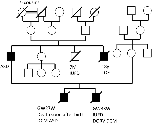
胎児期より拡張型心筋症と診断され周産期死亡した兄弟例Fetal Sibling Case of Familial Dilated Cardiomyopathy

1兵庫県立こども病院循環器科Department of Cardiology, Hyogo Children’s Hospital ◇ Hyogo, Japan

2日赤和歌山医療センター小児科Department of Pediatrics, Japanese Red Cross Wakayama Medical Center ◇ Wakayama, Japan

3加古川医療センター小児科Department of Pediatrics, Kakogawa Central City Hospital ◇ Hyogo, Japan

4富山大学医学部小児科Department of Pediatrics, Toyama University Hospital ◇ Toyama, Japan

5富山大学医学部法医学Department of Legal Medicine, Graduate School of Medicine University of Toyama Hospital ◇ Toyama, Japan

受付日:2017年10月19日Received: October 19, 2017
受理日:2017年12月21日Accepted: December 21, 2017
発行日:2018年1月1日Published: January 1, 2018
HTMLPDFEPUB3

胎児心エコーの進歩により,多くの先天性心疾患が出生前に診断されるようになったが胎児心筋症の報告は少ない.今回我々は,家族歴と胎児心エコーで拡張型心筋症(DCM)と診断し,周産期に死亡した兄弟例を経験した.父と父の弟がDCMで,両児とも顕微鏡受精で妊娠成立した.第1子は在胎21週に診断し,27週で母体Mirror症候群のため緊急帝王切開で出生したが,生後46分で死亡した.第2子は在胎27週に診断し,33週0日子宮内胎児死亡した.病理解剖所見はEndocardial elastofibrosis(EFE)で,家族性DCMと診断された.両児と両親の遺伝子解析では,父由来のTropomyosin α-1 chain(TPM1)遺伝子変異,母由来のdesmoplakin(DSP)遺伝子変異が同定された.家族性の胎児DCMで周産期死亡した場合,両親の遺伝子診断を行うことで次子の発症を予測できる可能性がある.

Fetal echocardiography is a well-established tool for prenatal diagnosis of many congenital heart diseases. However, there are few reports on the fetal diagnosis of cardiomyopathy. We herein report a case of siblings who were diagnosed as having familial dilated cardiomyopathy (DCM) by fetal echocardiography, pathological findings, and genetics analysis. Their father and father’s younger brother had DCM. Patient 1 was diagnosed at 21 weeks of gestation, and patient 2 at 27 weeks of gestation. Patient 1 was born by emergency cesarean section in the 27th week but died 46 minutes after birth. Patient 2 died in utero at 33 weeks of gestation. Pathological autopsy findings of both patients demonstrated endocardial elastofibrosis. Genetic analysis of both patients and their parents revealed a father-derived tropomyosin α-1 chain (TPM1) gene mutation and mother-derived desmoplakin (DSP) gene mutation. The prognosis of familial fetal DCM is poor, and careful family planning is necessary.

Key words: dilated cardiomyopathy; familial cardiomyopathy; fetal echocardiography; tropomyosin α-1 chain (TPM1) gene

はじめに

胎児心エコーの進歩により,多くの先天性心疾患が出生前に診断されるようになったが,胎児期に診断される心筋症の報告は少ない1–5).胎児心筋症の予後は,DCMでは不良で周産期死亡例が多く,特に妊娠中期から子宮内胎児死亡(IUFD)が起こり早期新生児死亡も多いといわれている1–5).American heart association(AHA)の心筋症分類によると,DCMは特発性混合型でそのうち家族性が約20~35%を占める6).特発性心筋症に関しては次世代シークエンサー等の遺伝子診断技術の発展により,DCM,肥大型心筋症(HCM),拘束型心筋症などの原因遺伝子の同定が進み,詳細な遺伝的背景が明らかになってきた.今回,我々は家族歴のある胎児DCMの兄弟例を経験した.両児とも周産期死亡後病理解剖を施行した.また,患児と両親の遺伝子診断を施行し,原因遺伝子と考えられる変異を同定した.今後,家族性DCMの出生前遺伝子診断の可能性を考えるうえで重要と思われるため報告する.

症例

第1子

母親38歳.0回経妊0回経産.既往歴は特になし.母親の家族歴は特になし.父親は2年前に職場の健康診断をきっかけにDCM,心房中隔欠損と診断された.父の同胞男児は在胎7か月で子宮内胎児死亡した.また,父の弟はファロー四徴で2歳,9歳時に手術既往があり,18歳時にDCMと診断され2か月後に死亡し病理解剖所見で左心室の拡大,心筋の錯綜配列と線維化を認めた.父方祖母は心臓肥大を指摘されたことがあるが,詳細は不明(Fig. 1).顕微鏡受精で妊娠成立し,前医で在胎13週より経過観察されていた.在胎21週の妊婦健診で胎児腹水,心房拡大を指摘され21週3日当科紹介受診となった.胎児心エコーで胎児心不全,三尖弁閉鎖不全,胎児心筋症疑い,左上大静脈遺残と診断した.この時点で生存出生の可能性が低く,IUFDの可能性があることを説明した.ご両親は妊娠の継続を希望された.在胎27週,母体Mirror症候群のため緊急帝王切開で出生したが生後46分で死亡した.染色体検査では正常核型であった.病理解剖を行った.

Pediatric Cardiology and Cardiac Surgery 34(1): 39-45 (2018)

Fig. 1 Pedigree of siblings with dilated cardiomyopathy (DCM) and their patients

Solid symbols indicate individuals with clinically defined DCM. Open symbols are unaffected members. Slashes indicate deceased members.

第2子

母親40歳.1回経妊1回経産.ヒトパルボウイルスB19 IgM0.28(−)IgG 8.08(+),サイトメガロウイルスIgM 0.60(−)IgG 2.0未満(−),抗SSA/Ro抗体7.0以下,抗SSB/La抗体7.0以下.凍結保存受精卵にて妊娠.第1子が周産期死亡したため妊娠管理目的にて在胎21週に当院産科紹介受診し,在胎21週5日に胎児心エコーを施行した.両大血管右室起始(DORV)と診断されたが心拡大なく心収縮は良好であった.在胎27週5日に胎児腹水のため,入院管理となり胎児心エコーを施行したところ両心室壁の菲薄化と心拡大,著明な壁運動の低下を認めDCMと判断した.ご両親の希望もあり,十分説明したうえで胎児心不全治療として母体にジゴシン投与したが,副作用が出現したため中止した.在胎33週0日にIUFDが確認された.翌日死産分娩し病理解剖を行った.

胎児心エコー所見

第1子では,在胎21週3日で胎児心拍数151bpm/分,総心横径(TCD)27.5 mm,心胸郭断面積比(CTAR)61%,三尖弁逆流(TR)中等度以上であった(Fig. 2A).臍帯静脈血流の逆流,心筋の非薄化と心拡大を認めDCMと判断した.

Pediatric Cardiology and Cardiac Surgery 34(1): 39-45 (2018)

Fig. 2 Fetal echocardiography of siblings with dilated cardiomyopathy

A: Fetal echocardiography of patient 1 at 21 weeks of gestation and 3 days showed a dilated LV and RV. RV; right ventricle, LV; left ventricle, Ao; aortic artery. B: Fetal echocardiography of patient 2 at 21 weeks of gestation and 5 days showed DORV and normal heart size without atrioventricular valve regurgitation. C: Fetal echocardiography of patient 2 at 27 weeks of gestation and 5 days showed a dilated LV and RV with tricuspid valve regurgitation (TR).

第2子では,在胎21週5日で胎児心拍数157 bpm/分,CTAR23%,TRなく心室中隔欠損と大動脈の騎乗を認めDORVと診断した(Fig. 2B).在胎27週5日では胎児心拍149 bpm/分,CTAR39%,TR中等度以上,臍帯静脈血流の逆流と心筋の非薄化,心拡大を認めた(Fig. 2C).Cardiovascular profile scoreは第1子4点,第2子が在胎21週10点,在胎27週4点であった(Table 1).

Table 1 Cardiovascular profile (CVP) score of the fetuses
Patient1 21W3dPatient2
21W5d27W5d
HydropsAscitesNoneAscites
Heart size(CTAR)61%23%39%
Cardiac functionHolosystolic TRNormalHolosystolic TR
Venous DopplerUV pulsatileNormal UV and DVUV pulsatile
Arterial DopplerUA retrogradeForward diastolic velocityForward diastolic velocity
CVP score4105
Patient 1 exhibits ascites, a dilated heart, tricuspid valve regurgitation, pulsatile pattern in the umbilical vein and retrograde flow in the umbilical artery. The CVP score of patient 1 was 4 points at 21 weeks of gestation. Patient 2 had normal cardiac function at 21 weeks of gestation. The CVP score of patient 2 was 10 points at 21 weeks of gestation. However, at 28 weeks of gestation, his CVP score decreased by 5 points.

病理解剖所見

第1子では組織像で心内膜の肥厚,弾性繊維の増殖および血管周囲性の繊維化を認めた(Fig. 3A, B).第2子では両心室の心内膜面は白色で左心室の壁厚は3.5 mm,右心室の壁厚は2.0 mmであった(Fig. 3C, D).組織像で心内膜肥厚は左心室および右心室にも及び(Fig. 3E),左心室心内膜弾性繊維の増生と血栓を認めた(Fig. 3F, G).第2子では,肉眼像で心臓は全体に丸みを帯び,心尖は鈍化していた(Fig. 4A).割面で心内膜はやや白色調であった(Fig. 4B).左心室;VSDがみられる.左室はやや低形成で内腔が拡張,心内膜面は白色調(Fig. 4C).壁厚は3.5 mm.右心室;ASDがみられる.心内膜面は全体的に白色調(Fig. 4D).壁厚は2 mm.組織像でHE染色では左心室心内膜肥厚,血栓,左心室心内膜弾性繊維の増生を認めた(Fig. 4A, B).Masson trichrom染色では左心室,右心室とも心内膜の肥厚,特に左心室の心内膜,弾性繊維増殖を認めた(Fig. 4C, D).診断は,第1子がASD PLSVC to CS, EFE疑いで,第2子がDORV VSD ASD, Hypoplasia and dilated of LV, Endocardial thickening with proliferation of elastic fiber LV and RV severe, EFE疑いであった.第1子,第2子とも家族歴,胎児心エコー所見,病理所見よりEFE,家族性胎児DCMと考えた.

Pediatric Cardiology and Cardiac Surgery 34(1): 39-45 (2018)

Fig. 3 Whole heart features and histological examination of brothers with dilated cardiomyopathy

A, B: Histological examination of patient 1. A: Endocardial thickening with proliferation of elastic fiber of LV. B: Perivascular fibrosis of LV. C–G: Macroscopic view and histological examination of patient 2. C: Cross-section through the LV showing diffuse white fibrous tissue covering the inner surface of the left ventricle. The LV cavity is dilated. D: Cross-section through the RV. The findings in the RV are similar to those in the LV. E: Endocardial hyperplasia also extended to both the RV and LV. F: Endocardial thickening with proliferation of elastic fibers and thrombus (arrows). G: Endomyocardial section showing proliferation of elastic fibers.

Pediatric Cardiology and Cardiac Surgery 34(1): 39-45 (2018)

Fig. 4 Results of genetic testing on the brothers and their parents

TPM1 mutation (c.712C>T, p. Srg238Trp) was detected in both patients and their father. They were diagnosed as having dilated cardiomyopathy. Desmoplakin gene mutation (c.4886G>T, p. Ser1629Ile) was detected in both patients and their mother. The mother was healthy and asymptomatic.

遺伝子診断

第2子の病理診断後に両児の心臓組織,両親の血液検体より遺伝子診断を行った.その結果,両児とも父由来のTPM1遺伝子変異(c.712C>T),母由来のDSP遺伝子変異(c.4886G>T)が同定された(Fig. 4).

考察

DCMは特発性混合性心筋症に分類され,そのうち20~35%が家族性である.木村らの解析では本邦での家族性DCM例の31%,孤発例DCM例の7%に遺伝子病変がみられる7).小児のDCM発症率は1/17~14万人8)と成人発症率に比べて低いが,乳児期発症例は4.4人/10万人と小児期発症に比べて高い9).小児DCMの予後は不良で心移植率および死亡率は5年で46%に達する9).遺伝性の要因が高く,家族性DCMの割合が成人の約2倍である10).小児DCMにおいて家族性DCMが予後不良因子であるという報告もあるが11),家族性と孤発性DCMの死亡率には差はないという報告もあり12)一定していない.

胎児心エコーの診断技術の発達によって,様々な先天性心疾患が胎児期に診断されるようになったが,胎児心筋症に関する報告は少ない1–5).胎児期DCMの発症率は不明であるが,Weberらは2002年から2012年まで年間約8万人の出生児中,61人に心筋症がみられたと報告した.このうち心筋肥大のない症例は40人で,10万人あたり約4.1人が発症しており,これは1歳以下での発症率と似通っている5).胎児症例ではTerminationの割合が多く,さらにIUFDと早期新生児死亡はあわせて82.3%と非常に高い1).胎児期に診断された家族性DCMの生存出生例は15~30%であった5)

Pedraらは家族性DCMの16家系26妊娠を調べたところ,8例で軽度から重症な心筋症を認めた2).同胞がDCMであった家系が11家系でそのうち18妊娠中7例が心筋症と診断された.胎児診断された4例は2例が胎児死亡し1例が早期産で出生後すぐに死亡している.この報告の他にいくつかの単施設での報告がみられるが,家族性DCMで胎児期に診断されている症例はほとんどが胎児死亡か新生児期早期死亡であった1, 3–5).小児期,成人期発症例と比べ,胎児期に発症した家族性DCMは非常に予後が悪いことが推察される.

CVPSは胎児水腫,静脈ドップラー波形,心拡大,心機能,動脈ドップラー波形という5項目を各々2点として計10点満点とし,低値であるほど胎児心不全を疑う指標である13).6点以下で予後不良といわれている.本報告の第2子では妊娠初期には問題なく中期以降に心機能低下,心拡大を示しCVPSも6点以下であった.同様の報告はいくつかあり,DCMの家族歴のある場合は妊娠中期にも胎児心エコー検査を行う必要がある2, 14)

家族性DCMの患者では30~40%にpathogenic variantが同定されるが,これらは各家系で個別に発生してその家系で受け継がれているprivate variantがほとんどで,一般に浸透率は低い15).本症例では父方由来のTPM1変異(c.712C>T)と母由来のDSP変異(c.4886G>T)が指摘された.TPM1はサルコメア蛋白の一種で肥大型心筋症(HCM)の原因遺伝子として知られているが,家族性DCMでも同定され2家系の報告がある.また,左室緻密化障害とDCMを合併する家系も報告されている.小児期に発症し予後不良の群と,成人発症した予後良好な群に分かれる.予後不良群では乳児突然死症例,心臓移植症例,心不全死症例が含まれるが一方で適切な治療により心機能が回復した症例もある16–18).組織所見は典型的なDCMであった16).今回見いだされた遺伝子変異は,新規の変異であり健常人での発症頻度とコンピューター解析から,障害性は明らかではないが父および父の兄弟にDCMとIUFDがみられることから,本症例に何らかの影響を与えていると考えられる(Table 2).一方,DSP変異は不整脈源性右室心筋症(ARVC)にみられるが,近年DCM症例でも報告されている19).母由来の遺伝子変異は正常人の0.76%にみられる変異であり病的意義の有無は不明であるが,周産期死亡した兄弟がともに同一の変異を有していたことから考えると,父方のTPM1変異に母方のDSP変異が加わることにより障害性が増した可能性が考えられる.ARVC, HCMでは複数の遺伝子変異が存在する場合,発症が早期で重症化するとされている20, 21).家族性DCMの心臓移植症例で複数以上の遺伝子変異を有する症例は21.2%とこれらの心筋症より高率で,幾つかの遺伝子変異が組み合わさって発症した家族性DCMの場合,症状はより重症になると推察されている22)

Table 2 Detailed descriptions of the genetic mutations
geneproteincodingCADDHGVDdbsnpSIFT (<0.05)polyPhen-2 (>0.9)
DSPp.Ser1629Ilec.4886G>T23.40.0076rs20024397600.454
TPM1p.Arg238Trpc.712C>T3301
CADD score: Combined annotation-dependent depletion is a tool for scoring the deleteriousness of single nucleotide variants. Both mutations had a high damage impact. Tropomyosin α-1 chain gene mutations are not detected in normal persons. HGVD: Human genetic variation database. This database is a reference of genetic variation in the Japanese population. The probability of genetic mutation of the desmoplakin gene in normal persons was 0.76%. dDbsnp: database of single nucleotide polymorphisms from the National Center of Biotechnology Information. Web source: NCBI dbsnp, SIFT, PolyPhen-2

本症例では次子も胎児期に致死性のDCMを発症する可能性がある.ご家族のさらなる挙児希望の意思を確認したため,他病院の遺伝子診療部を紹介し出生前診断もしくは着床前診断(preimplantation genetic diagnosis; PGD)の可否について検討していただいた.PDGは臨床研究として位置づけられ,対象疾患は重篤な遺伝子疾患に限られている.両親の少なくともいずれかに遺伝性素因があるために,その児に疾患が発症する可能性のある保因者に限って適応とすべきであるが,多くの遺伝性疾患の中で重篤性を一律に規定することは困難である23).家族性DCMの胎児期発症例の予後は不良であり,次子妊娠前に両親と児の遺伝子検査を行い原因遺伝子を同定できた場合,複数の遺伝子変異が存在すると,IUFDや新生児期早期死亡の可能性が高いことを示すことはできる.一方でDCMの予後は発症時期や原因により様々であり,致死性,重篤性を判断するのは難しい.症例の集積と家族の遺伝子診断を行うことにより,本症例のように周産期死亡が予想されるような場合における,PGDや出生前診断の可否に関して慎重に議論と検討を進めていく必要がある.

結語

今回,我々は周産期死亡したDCMの兄弟例を通して,病理診断と家族性DCMの遺伝子診断を行うことができた.家族性DCMで,胎児期にDCMを発症し周産期に死亡した場合,次子も同様の症状を呈する可能性がある.病理診断および遺伝子診断を行うことは,次子の発症の予測因子となる可能性があり有意義だと考える.

謝辞Acknowledgments

第1子,第2子の病理検査を施行していただきました国立循環器病センター病理部植田初江先生,遺伝カウンセリングを担当していただきました兵庫県立こども病院臨床遺伝科森貞直哉先生に深謝いたします.

利益相反

本論文について開示すべき利益相反(COI)はない.

付記

本論文の要旨の一部は第24回日本心筋疾患学術集会(2015年10月),第53回日本小児循環器病学会総会(2017年7月)において発表した.

引用文献References

1) Pedra SR, Smallhorn JF, Ryan G, et al: Fetal cardiomyopathies: Pathogenic mechanisms, hemodynamic findings, and clinical outcome. Circulation 2002; 106: 585–591

2) Pedra SR, Hornberger LK, Leal SM, et al: Cardiac function assessment in patients with family history of nonhypertrophic cardiomyopathy: A prenatal and postnatal study. Pediatr Cardiol 2005; 26: 543–552

3) Yinon Y, Yagel S, Hegesh J, et al: Fetal cardiomyopathy-in utero evaluation and clinical significance. Prenat Diagn 2007; 27: 23–28

4) Fesslova V, Mongiovì M, Pipitone S, et al: Features and outcomes in utero and after birth of fetuses with myocardial disease. Int J Pediatr 2010; 2010: Article ID 628451, 9 pages

5) Weber R, Kantor P, Chitayat D, et al: Spectrum and outcome of primary cardiomyopathies diagnosed during fetal life. JACC Heart Fail 2014; 2: 403–411

6) Maron BJ, Towbin JA, Thiene G, et al: Contemporary definitions and classification of the cardiomyopathies: An American Heart Association Scientific Statement from the Council on Clinical Cardiology, Heart Failure and Transplantation Committee; Quality of Care and Outcomes Research and Functional Genomics and Translational Biology Interdisciplinary Working Groups; and Council on Epidemiology and Prevention. Circulation 2006; 113: 1807–1816

7) 木村彰方:心筋症の分子病因・病態の解明から臨床へ.心臓2014; 46: 4–14

8) Lipshultz SE, Sleeper LA, Towbin JA, et al: The incidence of pediatric cardiomyopathy in two regions of the United States. N Engl J Med 2003; 348: 1647–1655

9) Towbin JA, Lowe AM, Colan SD, et al: Incidence, causes, and outcomes of dilated cardiomyopathy in children. JAMA 2006; 296: 1867–1876

10) Puggia I, Merlo M, Barbati G, et al: Natural history of dilated cardiomyopathy in children. J Am Heart Assoc 2016; 5: e003450

11) Alexander PM, Daubeney PE, Nugent AW, et al: Long-term outcomes of dilated cardiomyopathy diagnosed during childhood: Results from a national population-based study of childhood cardiomyopathy. Circulation 2013; 128: 2039–2046

12) Rusconi P, Wilkinson JD, Sleeper LA, et al: Differences in presentation and outcomes between children with familial dilated cardiomyopathy and children with idiopathic dilated cardiomyopathy: A report from the Pediatric Cardiomyopathy Registry Study Group. Circ Heart Fail 2017; 10: e002637

13) Huhta JC: Guidelines for the evaluation of heart failure in the fetus with or without hydrops. Pediatr Cardiol 2004; 25: 274–286

14) Barberato MF, Barberato SH: Foetal dilated cardiomyopathy with left ventricular thrombosis. Cardiol Young 2015; 25: 1191–1192

15) 森田啓行:【遺伝性心筋疾患の現状と展望を識る】識る 心機能異常を伴う遺伝性心筋疾患(解説/特集).Heart View 2016: 20; 151–156

16) Lakdawala NK, Dellefave L, Redwood CS, et al: Familial dilated cardiomyopathy caused by an alpha-tropomyosin mutation: The distinctive natural history of sarcomeric dilated cardiomyopathy. J Am Coll Cardiol 2010; 55: 320–329

17) Rampersaud E, Siegfried JD, Norton N, et al: Rare variant mutations identified in pediatric patients with dilated cardiomyopathy. Prog Pediatr Cardiol 2011; 31: 39–47

18) van de Meerakker JB, Christiaans I, Barnett P, et al: A novel alpha-tropomyosin mutation associates with dilated and non-compaction cardiomyopathy and diminishes actin binding. Biochim Biophys Acta 2013; 1833: 833–839

19) Elliott P, O’Mahony C, Syrris P, et al: Prevalence of desmosomal protein gene mutations in patients with dilated cardiomyopathy. Circ Cardiovasc Genet 2010; 3: 314–322

20) Quarta G, Muir A, Pantazis A, et al: Familial evaluation in arrhythmogenic right ventricular cardiomyopathy/clinical perspective: Impact of genetics and revised task force criteria. Circulation 2011; 123: 2701–2709

21) Garcia-Pavia P, Vázquez ME, Segovia J, et al: Genetic basis of end-stage hypertrophic cardiomyopathy. Eur J Heart Fail 2011; 13: 1193–1201

22) Cuenca S, Ruiz-Cano MJ, Gimeno-Blanes JR, et al: Genetic basis of familial dilated cardiomyopathy undergoing heart transplantation. J Heart Lung Transplant 2016; 35: 625–635

23) 末岡 浩:【生殖医療における倫理的問題を考える】着床前診断.産科と婦人科2016: 83; 297–302

This page was created on 2018-02-13T13:39:16.195+09:00
This page was last modified on 2018-03-06T11:32:05.452+09:00


このサイトは(株)国際文献社によって運用されています。