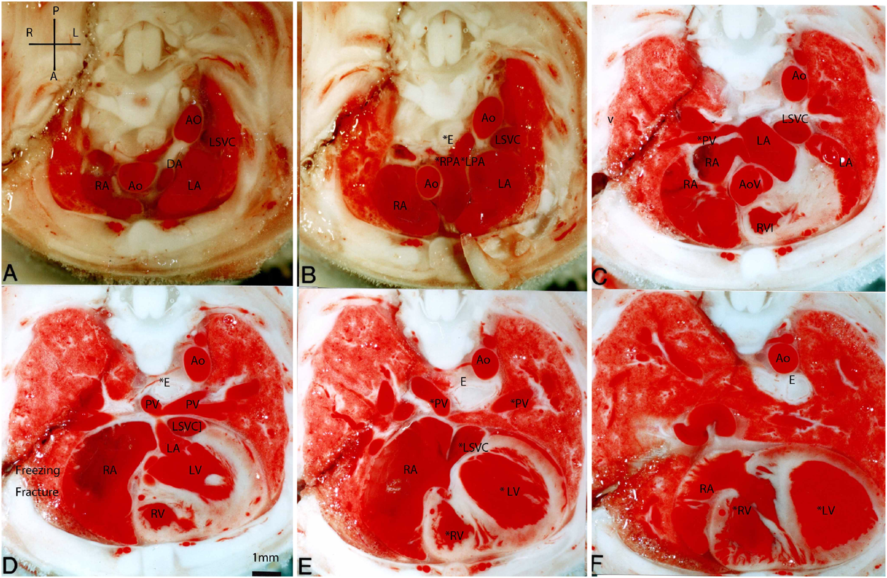
Online ISSN: 2187-2988 Print ISSN: 0911-1794
特定非営利活動法人日本小児循環器学会 Japanese Society of Pediatric Cardiology and Cardiac Surgery
Pediatric Cardiology and Cardiac Surgery 34(4): 165-171 (2018)
doi:10.9794/jspccs.34.165

ReviewReview

胎児循環器断面像卵円孔,静脈管,肺循環路の出生変化Fetal and Neonatal Cardiovascular Cross-Sectional Morphology in the Rat: The Foramen Ovale, Ductus Venosus, Right Ventricle, and Pulmonary Artery

東京女子医科大学循環器小児科Department of Pediatric Cardiology, Tokyo Women’s Medical University ◇ Tokyo, Japan

発行日:2018年12月20日Published: December 20, 2018
HTMLPDFEPUB3

本稿では胎児特有の循環路の出生時変化をラット凍結断面カラー写真の図譜で提示する.動脈管と先天性心疾患は紙面の都合で別の図譜にまとめた.ラット胎仔と新生仔を全身急速凍結法で固定し凍結ミクロトームで胸部を前額面,矢状面,横断面で切り,0.5 mm毎に断面カラー写真を連続撮影した.これにより胎児エコー図のモデルになる鮮明な天然色画像が得られた.心臓短軸断面で下大静脈右房開口部から卵円孔を経て左房への胎生期酸素化血の主要血流路が1断面に明示された.胸部矢状面でも臍静脈–静脈管–下大静脈–右房–卵円孔–左房の断面が描出された.新生仔の前額面では動脈管と卵円孔の閉鎖が明示された.胎仔で短い薄い卵円孔部心房中隔は生後2日で急速に進展肥厚して卵円孔を閉鎖した.胎生期心臓連続断面では太い動脈管,大きい胸腺,大きい右房と左房が明示された.いずれの断面図でも出生後に左右肺動脈末梢と肺静脈の急速な拡大,左右心室の拡大,右室壁菲薄化,肺の拡張,右心系のチアノーゼと左心系の動脈血化が明示された.

This review examines the natural-colored, cross-sectional morphology of the foramen ovale (FO), ductus venosus (DV), right ventricle (RV), and pulmonary artery (PA) in fetal and neonatal rats. The ductus arteriosus (DA) and congenital heart disease are reviewed separately. The in situ cross-sectional morphology of fetal and neonatal rat hearts was studied by rapid whole-body freezing, slicing on a freezing microtome, and serially photographing the cross-sections every 0.5 mm with a stereoscopic microscope (Wild M400). The serial frontal sections of the fetal thorax and upper abdomen showed the fetal oxygenated blood route including the umbilical vein, DV, inferior vena cava, posterior right atrium, FO, and left atrium. Neonatal frontal sections of the same area revealed closure of the DV, FO, and DA, in addition to rapid enlargement of the peripheral pulmonary vessels. Neonatal closure of the FO was associated with remarkable growth of the atrial septum flap. The fetal atrial septum was short and thin at the FO, and the flap tip only just reached the rim of the FO. Neonatally, this atrial septum grew rapidly in length and thickness within two days, closing the FO.

Key words: fetus; foramen ovale; ductus venosus; right ventricle; pulmonary artery

はじめに

小児循環器学で胎児心臓病学はますます重要になり,最近本誌に初めて胎児循環生理がReviewされた1).このReviewで明らかなのは胎児循環の生理はヒト胎児で研究することが困難なため,主に動物(羊)で研究2)されたことである.胎児循環の解剖も同様3)である.胎児循環は卵円孔,動脈管,静脈管など特有の循環路を持ち,これらは臨床上不鮮明ながら胎児エコーで観察4)されている.動物実験ではラットと全身急速凍結法を用いると胎生期循環路の鮮明な天然色の生体断面写真が得られる.この実験法はKarolinska研究所で開発され5),私は40年間動脈管の薬理学の研究6),心臓と大血管の出生時の形態変化の研究7–12),先天性心疾患の形態研究13–15),静脈管の研究11)に用いてきた.心臓断面画像はヒト胎児エコーのモデルにもなる.私はこれらの画像を白黒写真で発表したが,天然色断面画像の大部分は未発表なので,ここに図譜として発表する.なお胎生期先天性心疾患及び動脈管の断面像は別の論文14, 15)にまとめた.それぞれの実験の数字データは紙面の関係で省略するので,原著論文6–13)を参照されたい.

方法

ラット満期胎仔の全身急速凍結法による実験6–12)を次のように行った.ラットの妊娠期間は21.5日なので,妊娠21日目に親ラットを頸椎脱臼法で安楽死させ,ただちに帝王切開で取り出した胎仔を胎盤つきのままドライアイス−アセトン(−76°C)に投入して瞬時に凍結した.凍結した胸部を凍結ミクロトームで前額面frontal,矢状面sagittal,横断面transverse section,または心臓短軸面cardiac short sxisで切り,0.1乃至0.5 mm毎に実体顕微鏡(Wild M400)とカラーフィルム(Reale,富士フィルム)で撮影した.1個体の胸部から約20断面を撮影した.生後1~4日後のラットは予定日に自然出産後に親の保育授乳中のラットを用いた.

ラットの解剖学書16)によるとラットとヒトでは心臓と胸部大血管の解剖は大体共通しているが,次の種差がある.冠状静脈洞に還流する左上大静脈はヒトでは疾患として生じるがラットなどげっ歯類では正常である.左右の肺静脈はヒトでは胎生期後半以後は左右2本ずつ左房に入る17)が,ラットでは左房後方で合流して共通肺静脈になり,左房に接続する.臍動脈はヒトでは2本あるがラットでは1本である.臍静脈の腹壁から静脈管までの走行はヒトでは肝臓下面であり,ラットでは肝臓下面と横隔膜の中間で肝臓内である.

心臓血管断面写真

図中の重要な部位には*印を付した.

Fig. 1は胎仔と新生仔の卵円孔の前額面断面である.胎仔の卵円孔部の心房中隔flapは薄く,左房側に開いているが,生後2日で厚く長くなり,閉鎖する.左右肺動脈は生後急速に拡大する.

Pediatric Cardiology and Cardiac Surgery 34(4): 165-171 (2018)

Fig. 1 Frontal sections of the atrial septum of a rat fetus (A), a 1-day old rat (B), and a 2-day-old rat (C)

Abbreviations used in the figures. A: anterior. Ao: aorta. AoV: Aortic valve. Ca: caudal. Cr: cranial. DA: ductus arteriosus. DAo: descending aorta. DV: ductus venosus. E: esophagus. FO: foramen ovale. HV: hepatic vein. IAS: interatrial septum. IVC: inferior vena cava. L: left. LA: left atrium. LB: left bronchus. LPA: left pulmonary artery. LSVC: left superior vena cava. MPA: main pulmonary artery. p: posterior. PFO: patent foramen ovale. PoV: portal vein. PV: portal vein, pulmonary vein. PVa: pulmonary valve. LPA: left pulmonary artery. LPV: left pulmonary vein. R: right. RA: right atrium. RPA: right pulmonary artery. RV: right ventricle. RVI: right ventricular infundibulum. SVC: superior vena cava. T: trachea, thymus. US: umbilical sinus. UV: umbilical vein.

Fig. 2はラット胎仔心臓の短軸面断面を心室部(F)より順次心基部(肺動脈弁と大動脈弁部)(D)を経て主肺動脈部(C)大動脈弓部(B, A)へ切り撮影した.Cには胎仔の動脈血の血流路:下大静脈–右房後部–卵円孔–左房が明示されている.右室は左室室と同じ壁厚を持ち容積は左室よりやや大きい(E, F).左右肺動脈径は主肺動脈径の半分である.

Pediatric Cardiology and Cardiac Surgery 34(4): 165-171 (2018)

Fig. 2 Serial cardiac short-axis sections of a rat fetus

同じ所見はFig. 3でも見られる.Fig. 3は胎仔の心臓と肝臓の前額面図である.ほぼ同じ太さの動脈管,大動脈,肺動脈の断面像が示されている.臍静脈血が静脈管–卵円孔–左房へ流れる道筋14)が(F)に示されている.細めの左右肺動脈が(G)に明示されている.

Pediatric Cardiology and Cardiac Surgery 34(4): 165-171 (2018)

Fig. 3 Serial frontal sections of the heart and liver of a normal rat fetus. The final panel shows a sagittal section indicating the section planes for sections (A) to (I)

Fig. 4は生後1日の新生仔心臓後部と肝臓の前額面,Fig. 5は生後4日の新生仔である.静脈管3)は閉鎖し,左右肺動脈の急速な生後の拡大7)がある.ただし左右肺動脈起始部(Fig. 4A, Fig. 5A)は特に左肺動脈で末梢部より細い7)

Pediatric Cardiology and Cardiac Surgery 34(4): 165-171 (2018)

Fig. 4 Serial frontal sections of the posterior heart and liver of a 1-day-old rat

Pediatric Cardiology and Cardiac Surgery 34(4): 165-171 (2018)

Fig. 5 Serial frontal sections of the posterior heart of a 4-day-old rat

Fig. 6Fig. 7はラット胎仔の臍静脈,静脈管7, 11)を示す.ヒトの静脈管は腹壁から肝臓下面を背中方向へ走行するが,ラットの臍静脈3)は前腹壁から肝臓内を背中方向へ走行する.

Pediatric Cardiology and Cardiac Surgery 34(4): 165-171 (2018)

Fig. 6 Serial sagittal sections of the umbilical vein, umbilical sinus, portal vein, ductus venosus, and inferior vena cava of a rat fetus. The cutting planes are shown in E

Pediatric Cardiology and Cardiac Surgery 34(4): 165-171 (2018)

Fig. 7 Serial frontal sections of the fetal heart and liver showing the open fetal channels: the ductus arteriosus, right and left pulmonary arteries, umbilical sinus, ductus venosus, and foramen ovale. The cutting planes are shown in the upper right corner

Fig. 8Fig. 9はラット胎仔と新生仔(生後2日)の胸部横断面である.新生仔では胎仔のチアノーゼがなくなり,動脈管は閉じ,肺は拡張し,右房と左房はやや小さく,右室と左室は拡張し,右室壁は薄い10)

Pediatric Cardiology and Cardiac Surgery 34(4): 165-171 (2018)

Fig. 8 Serial thoracic transverse sections of a normal rat fetus

Pediatric Cardiology and Cardiac Surgery 34(4): 165-171 (2018)

Fig. 9 Serial thoracic transverse sections of a 2-day-old rat

おわりに

臨床で新生児の心エコーをとると,小さい心房中隔欠損がしばしば見つかるが,ここに提示したラットでの断面図から卵円孔自然閉鎖機転は明らかで,この種の心房中隔欠損の良性の予後が理解される.新生児の機能性雑音の一つが左肺動脈起始部狭窄から生じ,ラット新生仔の肺動脈断面像に狭窄がでている.ラットの胎生期間は7日でヒトの7か月に相当し,胎仔と新生仔の成長はヒトより数十倍速やかであり,循環器の発達と変化も速やかである.

利益相反

利益相反に関して記載すべき事項はありません.

引用文献References

1) 稲村 昇:胎児循環生理.日小児循環器会誌2016; 32: 451–461

2) Rudolph AM: Congenital Diseases of the Heart: Clinical-Physiological Considerations. Third Ed., Chichester, UK, Wiley-Blackwell, 2009, pp 1–24

3) Polin RA, Fox WW, Abman SH: Fetal and Neonatal Physiology. Fourth Ed., Philadelphia, Elsevier, 2011

4) Yagel S, Silverman NH, Genbruch U (eds): Fetal Cardiology. Martin Denitz, London, 2003

5) Hörnblad PY, Larson KS: Studies on closure of the ductus arteriosus 1: Whole-body freezing as improvement of fixation proceedures. Cardiologia 1967; 51: 231–241

6) 門間和夫:動脈管薬の実験40年.日小児循環器会誌2016; 32: 261–269

7) Momma K, Ito T, Ando M: In situ morphology of the ductus venosus and related vessels in the fetal and neonatal rat. Pediatr Res 1992; 32: 386–389

8) Momma K, Ito T, Ando M: In situ morphology of the aorta and common iliac artery in the fetal and neonatal rat. Pediatr Res 1993; 33: 302–306

9) Momma K, Ito T, Mori Y, et al: In situ pulmonary vascular morphology and lung volume in the fetal and neonatal rat. Early Hum Dev 1993; 34: 191–198

10) Momma K, Takao A, Ito R, et al: In situ morphology of the heart and great vessels in fetal and neonatal rats. Pediatr Res 1987; 22: 573–580

11) Takeuchi D, Momma K: Effect of decreased umbilical blood flow and hemorrhage, and decreased prostaglandins on the ductus venosus diameter in the rat. Biol Neonate 2006; 89: 42–49

12) 門間和夫:出生時の循環器系の適応生理.小児科2000; 41; 2225–2231

13) Momma K, Ando M, Takao A: Fetal cardiac morphology of tetralogy of Fallot with absent pulmonary valve in the rat. Circulation 1990; 82: 1343–1351

14) 門間和夫:ラット胎仔先天性心疾患の断面像:胎児心エコーのための染色体22q11欠失症候群モデル動物図譜として.日小児循環器会誌2018; 34 : 55-62

15) 門間和夫:動脈管の収縮形態:ラット動脈管断面像天然色図譜.日小児循環器会誌2018; 34: 128–134

16) Hebel R, Stromberg MW: Anatomy and Embryology of the Laboratory Rat. Wörthsee, BioMed Verlag, 1986, pp 112–113

17) Anderson RH, Brown NA, Webb S: Development and structure of the atrial septum. Heart 2002; 88: 104–110

This page was created on 2018-08-13T14:37:20.808+09:00
This page was last modified on 2018-12-17T11:12:22.000+09:00


このサイトは(株)国際文献社によって運用されています。