This report describes the case of a 6-year-old male patient with a pericardial cyst. He was originally referred to our hospital for treatment of idiopathic pulmonary arterial hypertension, without any structural abnormalities of the heart.
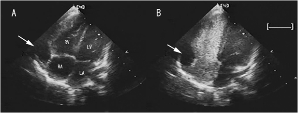
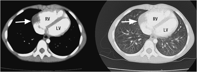
An echocardiographic study identified an anechoic lesion with a clear border located outside the right atrium (Fig. 1A). Chest contrast-enhanced computed tomography revealed a 16×14-mm lesion with the CT number of water (Fig. 2). A pericardial cyst or diverticulum of the right atrium was suspected, given the location and distinct visual features of the lesion.
The presence of a cyst was finally confirmed using microbubble echocardiography; this study was added to preplanned cardiac catheterization to evaluate pulmonary arterial hypertension. In this test, the contrast agent injected from the inferior vena cava did not flow into the cavity (Fig. 1B), indicating the lack of communication between the cavity and cardiac structures.
Pericardial cysts are rare mediastinal lesions; they are frequently located at the right cardiophrenic angle. The incidence rate is 1 in 100,000.1) Patients are usually asymptomatic, and the cysts are found incidentally on chest radiographs or echocardiography. They are mostly caused by the failure of fetal lacunae to coalesce into the pericardial coelom. However, the cysts can also occur as sequelae of pericarditis. Our patient had no history of pericarditis; therefore, the cyst was congenital. No etiological relationship was found between the cyst and the idiopathic pulmonary arterial hypertension.
We performed contrast echocardiography to distinguish the cyst from a diverticulum. If the anechoic lesion was a diverticulum, the chamber would be filled with contrast agent, as reported previously.2) Direct injection from the inferior vena cava yielded a vivid image, even with a small amount of contrast medium.
Pericardial cysts are usually benign and do not require surgical resection, which is only considered for symptomatic patients with chest pain or dyspnea. The cyst in our case appeared the same over one year and the patient showed no associated symptoms. However, we plan to continue conservative follow up, because some case reports describe cyst rupture, erosion of the cyst into the right ventricle or superior vena cava, and cardiac tamponade.3)
Conflicts of interest
None.
Note
Supplementary movies are provided online for this article.