Online ISSN: 2187-2988 Print ISSN: 0911-1794
特定非営利活動法人日本小児循環器学会 Japanese Society of Pediatric Cardiology and Cardiac Surgery
Pediatric Cardiology and Cardiac Surgery 32(1): 62-63 (2016)
doi:10.9794/jspccs.32.62

Images in Pediatric and Congenital Heart DiseaseImages in Pediatric and Congenital Heart Disease

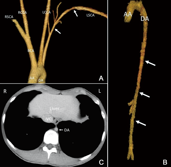
Stenoses in the Left Subclavian Artery and Descending Aorta in a Patient with Williams Syndrome

1Department of Pediatrics, Hokusetsu General Hospital ◇ Osaka, Japan

2Department of Radiology, Hokusetsu General Hospital ◇ Osaka, Japan

3Department of Pediatrics, Osaka Medical College ◇ Osaka, Japan

受付日:2015年9月3日Received: September 3, 2015
受理日:2015年11月9日Accepted: November 9, 2015
発行日:2016年1月1日Published: January 1, 2016
HTMLPDFEPUB3

Key words: Williams syndrome; Contrast-enhanced computed tomography; Arteriopathy

Pediatric Cardiology and Cardiac Surgery 32(1): 62-63 (2016)

Fig. 1 Contrast-enhanced computed tomography images of the aorta

(A) A contrast-enhanced computed tomography scan showing the aortic arch and its branches. Diffuse stenosis can be observed in the left subclavian artery (arrows). The sizes of the left and right subclavian arteries are compared. (AA, ascending aorta; BCA, brachiocephalic artery; RSCA, right subclavian artery; RCCA, right common carotid artery; LCCA, left common carotid artery; LSCA, left subclavian artery; DA, descending aorta). (B) A contrast-enhanced computed tomography scan showing the entire aorta. Diffuse and long stenosis can be observed in the entire descending aorta (arrows). The diameter of the ascending aorta (24.9 mm) is normal (mean normal diameter, 23.0 mm),3) whereas the diameter of the descending aorta (8.2 mm) is significantly smaller than the mean normal diameter (17.1 mm).3) (AA, ascending aorta; DA, descending aorta). (C) A contrast-enhanced computed tomography scan (axial view) at the lower thoracic level showing no thickening of the aortic wall. (DA, descending aorta; IVC, inferior vena cava) (R, right; L, left indicate the orientation.)

A 16-year-old boy with Williams syndrome, previously diagnosed by using fluorescence in situ hybridization, was observed to have different pulse intensities in his radial arteries, with the left radial artery pulse being weaker than the right radial artery pulse (blood pressures in the right and left brachial arteries, 128/66 and 116/58 mmHg, respectively). He previously presented with hypertension (blood pressure in the right brachial artery, 156/80 mmHg), which was effectively treated and controlled with candesartan (blood pressure in the right brachial artery, 130/88 mmHg). His hypertension was considered to have been caused by arteriopathy and not by renal hypoperfusion because no stenosis was observed in the renal arteries on abdominal ultrasonography (data not shown) and his plasma renin activity (2.1 ng/[mL·h]) was within the normal range (0.2–2.7 ng/[mL·h]). At first, magnetic resonance imaging (MRI) was considered, but because of his inability to cooperate in examinations that require a long time, contrast-enhanced computed tomography (CT), which requires a much shorter time, was performed instead. Contrast-enhanced CT of the aorta and subclavian arteries was performed, revealing diffuse narrowing of the left subclavian artery (Fig. 1(A)) and the entire descending aorta (Fig. 1(B)). There was no thickening of the aortic wall (Fig. 1(C)).No stenosis was observed in the supravalvular aortic region or the pulmonary arteries (data not shown).

In Williams syndrome, haploinsufficiency due to a deletion of chromosome 7q11.23, which involves the elastin gene, ELN, is implicated.1) Elastin arteriopathy is generalized and may affect virtually any artery. Arteriopathy frequently occurs; therefore, monitoring patients for arteriopathy and hypertension is important.2) To evaluate the status of systemic arteries, MRI is desirable because it confers no radiation exposure. However, if the patient tends to be uncooperative in examinations that require a long time and a wide range of systemic arteries need to be examined, contrast-enhanced CT is helpful in the evaluation of possible stenosis in the aorta and other arteries because it is noninvasive and easy to perform, especially in an outpatient setting.

This page was created on 2015-11-11T14:08:11.384+09:00
This page was last modified on 2016-01-26T14:05:10.62+09:00


このサイトは(株)国際文献社によって運用されています。