Online ISSN: 2187-2988 Print ISSN: 0911-1794
特定非営利活動法人日本小児循環器学会 Japanese Society of Pediatric Cardiology and Cardiac Surgery
Pediatric Cardiology and Cardiac Surgery 35(1): 9-17 (2019)
doi:10.9794/jspccs.35.9

ReviewReview

QT短縮とQT短縮症候群Short QT Interval and Short QT Syndrome

新潟大学医歯学総合病院魚沼地域医療教育センターUonuma Institute of Community Medicine, Niigata University Medical and Dental Hospital ◇ Niigata, Japan

受付日:2018年5月23日Received: May 23, 2018
受理日:2018年11月12日Accepted: November 12, 2018
発行日:2019年3月1日Published: March 1, 2019
HTMLPDFEPUB3

QT短縮症候群(short QT syndrome: SQTS)は遺伝性の致死的不整脈である.2000年に初めて報告され,心電図上のQT短縮を特徴とし,心室細動や心房細動などの不整脈を来す.臨床像は未だ不明な点も多いが,10~20歳台の突然死が多く報告されている.よって小児期での診断が重要であり,学校心臓検診でも問題になる.2013年の三大陸合同のExpert consensus statement以降は,QTcが一定基準以下のものは,遺伝子異常も家族歴もない無症状例もSQTSと診断される.一般成人集団よりQT短縮例を抽出する検討では,SQTSの診断基準を満たす例はあったが無症状で予後も良いと報告されている.我々の学校心臓検診の検討でもSQTSは存在したが,有症状例はなかった.SQTSの中でも高リスクの既報例と検診・健診などで抽出される低リスク例には臨床像に大きな隔たりがあり,SQTSのリスク層別化が重要となる.そこで我々は,有症状のSQTS既報例と学校心臓検診で抽出された無症状で家族歴もないQT短縮例の心電図を比較し,高リスクのSQTS例の鑑別が可能か検討した.さらにSQTSのリスク層別化を進めるには無症状のSQTS小児例の中長期予後を知ることが求められる.

Short QT syndrome (SQTS) is a hereditary lethal arrhythmia; it was first described in 2000. SQTS is characterized by an abnormal short QT interval on electrocardiogram (ECG), and causes arrhythmia, such as ventricular fibrillation and atrial fibrillation. Although clinical characteristics of SQTS have not been appropriately elucidated, several victims, individuals in the teenage and those in their 20s, have been reported. Therefore, SQTS diagnosis at an early age is important and is recognized as a concern in a school screening program for heart disease. According to an expert consensus statement published in 2013, even an asymptomatic individual with a short QT interval, who has neither a family history nor a pathogenic gene mutation, can be diagnosed with SQTS. Studies with various adult populations have identified individuals who met the SQTS diagnostic criteria; however, these patients were asymptomatic and did not have a poor prognosis. Some asymptomatic children and adolescents with SQTS were also identified in our study, in which we mainly focused on a school screening program for heart disease. Risk stratification of SQTS is necessary because clinical characteristics of SQTS are variable, including no symptoms and sudden cardiac death. Hence, we compared the ECGs of asymptomatic individuals with a short QT interval in a school screening program for heart disease with those of previously reported symptomatic patients with SQTS, to screen for SQTS patients with a high risk. A prospective study thus needs to be conducted to elucidate the prognosis of asymptomatic patients with SQTS who are diagnosed in their childhood.

Key words: short QT syndrome; QT interval; sudden cardiac death; lethal arrhythmia

はじめに

QT短縮症候群(short QT syndrome: SQTS)は心電図上のQT短縮を特徴とし,心室細動や心房細動を発症する.10~20歳台の突然死が多く報告され,小児期での診断が重要である.近年,3大陸合同のExpert consensus statement1)や欧州心臓病学会2)から発表された診断基準では心電図上のQT短縮所見のみでもSQTSと診断される.無症状から突然死まで臨床像は幅広く,リスク層別化が求められる.学校心臓検診でも,家族歴のない無症状のSQTS例が抽出され,その扱いが問題になっている.

本稿では,まず診断の注意点や臨床像などSQTS全般について述べ,次に学校心臓検診など,集団より抽出されるQT短縮例(SQTSを含む)の頻度や特徴,さらにSQTSのリスク層別化について述べる.

SQTSとは

SQTSは心電図でのQT短縮が特徴の致死的不整脈である.2000年に初めて報告され3),現在までに約100例の症例報告がある.心臓の形態異常は伴わない.原因としてイオンチャネル遺伝子異常が知られ,6種類の責任遺伝子が報告されているが4)Fig.1),遺伝子異常が同定されない症例が半数以上ある(Fig. 2).心室頻拍や心室細動,心房細動が惹起され,失神や若年突然死の原因となる.臨床像はまだ不明な点が多い.

Pediatric Cardiology and Cardiac Surgery 35(1): 9-17 (2019)

Fig. 1 Mechanism of SQTS

(a) Electrocardiogram in a normal subject (upper panel) and a patient with SQTS (lower panel). The QT interval is abnormally shortened in a patient with SQTS. (b) Contributions of ionic currents to an action potential in a ventricular cardiomyocyte in a normal subject (upper panel) and a patient with SQTS (lower panel). Red and blue arrows indicate inward and outward currents, respectively. In a patient with SQTS, an abnormal decrease in the inward current or an abnormal increase in the outward current causes shortening of the action potential duration. In SQT1, SQT2, SQT3, and SQT4-6, IKr, IKs, IK1, and ICaL are affected, respectively. Double headed arrows indicate an allelic relationship. (c) Genes causing SQTS. IK1, background potassium; INa, fast inward sodium current; Ito, early transient outward potassium current; ICa, inward calcium current; IKr, rapid delayed rectifier potassium current; IKs, slow delayed rectifier potassium current; LQT1, 2, 7, and 8 indicate long QT syndrome types 1, 2, 7, and 8, respectively.

Pediatric Cardiology and Cardiac Surgery 35(1): 9-17 (2019)

Fig. 2 Genetic diagnosis of the reported cases with SQTS

The group of “genetic diagnosis (−)” comprised patients in whom the genetic test was not performed, indicated negative or was not reported.

1. SQTSの病態生理

イオンチャネル異常により,内向きカルシウム電流の減弱や外向きカリウム電流の増強が起こり,再分極が早まる.心筋細胞レベルでは活動電位は短縮し,心電図上ではQT間隔が短縮する(Fig. 1).不均一な再分極に伴うphase 2 reentryが心室細動や心室頻拍の引き金と考えられ,QT延長症候群に似る5)

2. SQTSの診断

1)診断基準

2011年にGollobらが初めてSQTSの診断基準を提唱した6).QT延長症候群のSchwartzスコアのようにスコア化されている.QT間隔に加え,Jpoint-Tpeak間隔,既往歴,家族歴,遺伝子検査の所見を加味し,診断する(Table 1).その後,診断基準は2013年に3大陸合同のExpert consensus statementで発表され1)Table 2),2015年に欧州心臓病学会のガイドラインで示された2)Table 3).この2つではQT短縮所見のみでもSQTSと診断しうる.Expert consensus statementではQTc≦330 ms,欧州心臓病学会ではQTc≦340 msがSQTSとなる.この点以外は2つの診断基準はほぼ同じである.いずれの診断基準でもBazettの心拍補正を用いている.

Table 1 SQTS diagnostic criteria presented by Gollob et al.6)
Point
QTc (Bazett) ms
<3701
<3502
<3303
Jpoint-Tpeak interval<120 ms1
Clinical history
History of sudden cardiac arrest2
Documented polymorphic VT or VF2
Unexplained syncope1
Atrial fibrillation1
Family history
1st or 2nd -degree relative with high-probability SQTS2
1st or 2nd -degree relative with autopsy-negative SCD1
SIDS1
Genotype
Genotype positive2
Mutation of undetermined significance in a culprit gene1
High-probability SQTS: ≥4 points, intermediate-probability SQTS: 3 points, low-probability SQTS: ≤2 points. Electrocardiogram: must be recorded in the absence of modifiers known to shorten the QT interval. J point-T peak interval must be measured in the precordial lead with the greatest amplitude T-wave. Clinical history: events must occur in the absence of an identifiable etiology, including structural heart disease. Points can only be received for 1 of cardiac arrest, documented. polymorphic VT, or unexplained syncope. Family history: points can only be received once in this section. * A minimum of 1 point must be obtained in the electrocardiographic section in order to obtain additional points. VF; ventricular fibrillation, VT; ventricular tachycardia
Table 2 Expert consensus recommendations on SQTS diagnosis1)
SQTS is diagnosed;
QTc (Bazett)≦330 ms
SQTS can be diagnosed;
QTc (Bazett)360 ms
and one or more of the following:
① a pathogenic mutation
② family history of SQTS
③ family history of sudden death at age 40 years
④ survival of a VT/VF episode in the absence of heart disease
Red font indicates differences from the European Society of Cardiology guideline (Table 3).
Table 3 Diagnosis of SQTS according to the European Society of Cardiology guideline2)
SQTS is diagnosed;
QTc (Bazett)≦340 ms
SQTS should be diagnosed;
QTc (Bazett)360 ms
and one or more of the following:
① a confirmed pathogenic mutation
② family history of SQTS
③ family history of sudden death at age40 years
④ survival of a VT/VF episode in the absence of heart disease
Red font indicates differences from expert consensus recommendations (Table 2).
2)診断時の注意点

診断にはいくつかの注意点がある.まず2次性のQT短縮を除外しなければならない.様々な原因でQT短縮が起こる7)Table 4).

Table 4 Secondary causes of short QT interval
Hyperkalemia
Hypercalcemia
Hyperthermia
Acidosis
Effect of catecholamine
Activation of I K Ach
Activation of I K ATP
Effects of drugs such of digitalis
Carnitine deficiency
Modified based on reference 7.

QT間隔の測定法や心拍補正法によりQTcは異なる8).Bazett補正では徐脈時はQT間隔を過剰補正してSQTSを過剰診断,頻脈時にはQT間隔を過小補正してSQTSを過小診断する懸念がある.一方Fridericia補正では頻脈時に過剰診断する懸念がある.また頻脈時には正常者とQT間隔がoverlapする可能性ある9).このために心拍数60/分前後での評価が推奨されている.しかし小児では心拍数60/分前後での心電図測定が困難なことが多いのが問題である.

さらに洞機能不全例での評価には注意を要する.洞機能不全があると交感神経刺激によりQT間隔が短縮しても,心拍数上昇(RR間隔短縮)が伴わないためQTcが短縮する可能性がある10)

3. SQTS既報例の臨床像

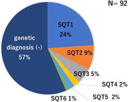
過去のSQTS患者についての報告で,1)有症状,2)SQTSまたは若年突然死の家族歴,3)遺伝子診断確定,のうち少なくとも一つを満す既報例を対象として,臨床像をまとめた4, 6, 11–24).対象患者は92例だった.Bazett補正QTcは194~377 msであった.うちQTc>360の2例は遺伝子診断され,突然死の家族歴があった6).それ以外の症例は3大陸合同のExpert consensus statementの診断基準を満たしていた.約6割が遺伝子型不明であった(Fig. 2).

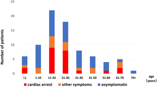
男性が多く,男女比は7 : 3であった.診断時年齢は0~80歳と幅広いが,30歳以下の若年者が3分2以上を占めた(Fig. 3).心停止例は92例中25例(27%)で,うち30歳以下が8割以上を占め,若年者がほとんどであった(Fig. 3).発症年齢に性差があった(Fig. 4).男性では10~20歳代での発症が殆どだが,女性では発症年齢の偏りはなかった.思春期以降の男性に発症する点はBrugada症候群に似ており,男性ホルモンの影響が推察される.しかし発症年齢は30~40歳代が多いBrugada症候群に比しSQTSは少し若い.その明確な理由はわからないが,これはBrugada症候群と比べ加齢の影響が少ないのかもしれない.

Pediatric Cardiology and Cardiac Surgery 35(1): 9-17 (2019)

Fig. 3 Age distribution of onset or diagnosis of the reported cases with SQTS

Age at the time of diagnosis is shown in asymptomatic patients with SQTS.

Pediatric Cardiology and Cardiac Surgery 35(1): 9-17 (2019)

Fig. 4 Age distribution according to sex in the onset of the reported cases with SQTS

心房細動は17例(18.5%)に,洞機能不全は7例(7.6%)に合併していた.心房細動はSQT1−4に報告があった.一方,洞機能不全合併7例中6例は胎児期徐脈を契機に診断されたSQT2,KCNQ1-V141M変異例であった.この変異は胎児新生児期の徐脈と若年発症心房細動が主体である点,QT短縮を伴わない洞機能不全例や心電図異常を認めない例もあり,型に幅がある点など,他のSQTSとは臨床像が異なった.

無症状例は47例(51%)であった.

4. 検査

1)12誘導心電図

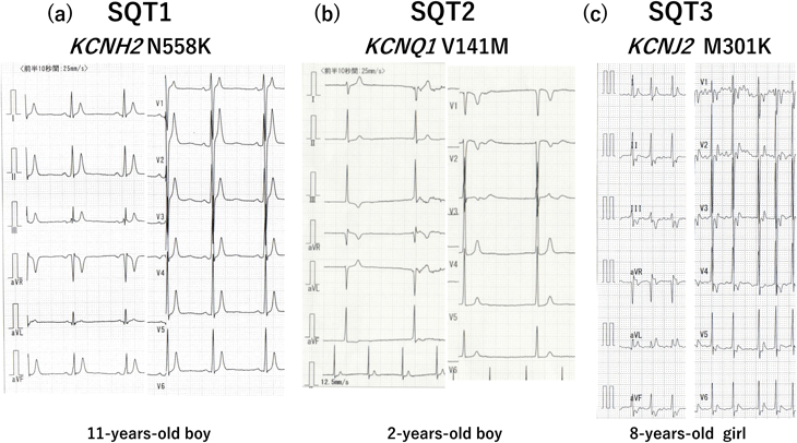
診断に最も重要である.QT間隔を評価するだけでなく,T波形も重要な所見である.T波形は遺伝子型で異なることが知られている(Fig. 5).SQT1では高く左右対称性なT波形が多い.最も報告の多いKCNH2-N588K変異13, 25)や他の変異でも報告されている12).SQT2は様々である.最も報告の多いKCNQ1-V141MのT波は高くないが24),高いT波を示す他の変異の報告もある14).SQT3では左右非対称なT波の報告が多い.T波が急峻に立ち上がる例と17),逆にTpeakから急峻に下降する例がある6).SQT4-5はBrugada型心電図を示す23).QT短縮の程度と重症度の関連は示されていない.12誘導ECGの所見によるリスク層別化の試みは後述する.

Pediatric Cardiology and Cardiac Surgery 35(1): 9-17 (2019)

Fig. 5 Twelve-lead electrocardiograms of SQT1, 2, and 3

(a) SQT1; T waves are narrow, tall, and peaked in our case. (b) SQT2; T waves are neither tall nor peaked (data were kindly provided by Dr. Tateno, Chiba Cerebral and Cardiovascular Center, Ichihara, Japan). (c) SQT2; T waves are asymmetrical with a rapid ascending phase (data were kindly provided by Dr. Ehara, Osaka City Hospital, Osaka, Japan).

2)運動負荷試験・ホルター心電図

運動負荷試験やホルター心電図においてはQT間隔が短縮し,正常に比し心拍数の変化に伴うQT間隔の変化が乏しいのが特徴である9).ホルター心電図は心房細動の合併評価にも有用である.

3)電気生理学検査

心室細動や心室頻拍の誘発頻度は低いが,心室不応期は短い.正常群と比較した検討では,右室流出路の不応期200 ms未満を基準とすると感度86%,特異度100%と報告がある26)

5. 生活管理と治療

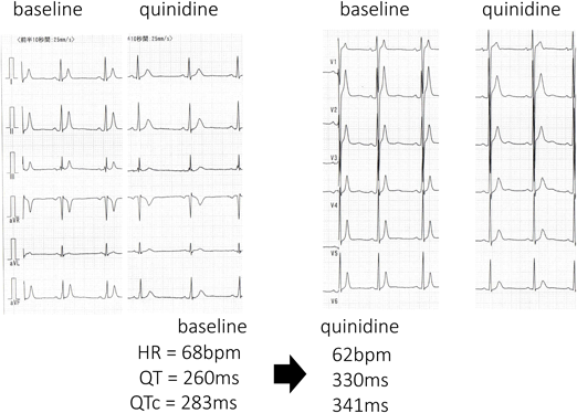
生活管理や治療に関するエビデンスは乏しく,運動制限の有効性は示されていない.薬物治療も十分と言えるエビデンスはないが,キニジンの有効性が報告されている.特にSQT1でQT間隔が正常化する例があり心イベント抑制効果が期待される25, 27)Fig. 6).ソタロールなどのIII群薬はSQT1に対するQT延長効果は乏しいが,他のサブタイプで効果が期待されている.Expert consensus statement1)や欧州心臓病学会のガイドライン2)では,「突然死の家族歴のある無症状例に対してキニジンやソタロール投与を考慮しても良いかもしれない」としている(class IIb).

Pediatric Cardiology and Cardiac Surgery 35(1): 9-17 (2019)

Fig. 6 Electrocardiogram showing the effects of quinidine (20 mg/kg/day) in an 11-year-old boy with SQT1

HR, heart rate; QTcB, the QT interval corrected by Bazett’s formula

ICDが突然死防止に有用と言えるが,QTが極端に短縮しているため,T波をQRSと認識し誤作動する場合がある.Expert consensus statement1)と欧州心臓病学会2)のガイドラインではICD植え込みを,1)心停止既往例,2)持続性心室頻拍の既往者(意識の有無にかかわらず)に推奨している(class I).さらに突然死の家族歴のある無症状例については,Expert consensus statement1)では考慮してもよいかもしれないとし(class IIb),欧州心臓病学会のガイドライン2)では個々の症例での判断を勧めている.

集団より抽出されるQT短縮例

これまで様々な集団からQT短縮例を抽出した報告がある8, 10, 28–34).このなかにはSQTSの診断基準を満たす症例も含まれていおり,SQTSの頻度や臨床像を知ることにもつながる.

報告によりQT短縮例の頻度は異なり,QT間隔の測定方法の違いに加え,母集団の違いが頻度に影響していると推察される.成人の報告では人種,性別,年齢により頻度が異なると指摘している.男性,黒人にQT短縮例が多く,性ホルモン(テストステロン)の影響が推察されている33).病院受診例を対象とした報告では,男女共に10~20歳代にかけてQT短縮例が多かったが,その理由は不明である32).さらに競技スポーツ者にQT短縮例が多かったという報告があり,運動による再分極の変化が推察されている33).また成人QT短縮例の長期予後は良好と報告されている28, 29, 33).

小児の報告はわずかであるが,シンシナティ小児病院の心電図データベースを基にした報告がある34).21歳未満の心電図272,504件を対象としてBazett補正でQTc<340 msを抽出基準とし,45例(0.05%)が抽出された.平均10.7か月フォローし,2例が死亡したがいずれも不整脈死ではなかった.

一方,我々は一般小児のQT短縮の頻度を知るため,新潟市学校心臓検診の小学校1年生(小1)6,607名と中学校1年生(中1)6,707名を対象として研究を行った.QTc<330 msを抽出基準とした.抽出例数は,Bazett補正で小1は男1名(0.01%)のみ,中1は13名(0.2%,男9名)であった.またFridericia補正では小1は20名(0.3%,男7名),中1は11名(0.16%,男6名)であった.我々の一般小児集団が米国小児病院受診者集団より低い抽出基準であったのにもかかわらずQT短縮例が高頻度であった.どちらの報告にも有症状のSQTS例はなかった.

以上より,様々な集団から抽出されるSQTSのほとんどが無症状のようである.

SQTSのリスク層別化

SQTSのなかでも,高リスクの既報例と検診・健診などで抽出される低リスク例には臨床像に大きな隔たりがあり,高リスク例の鑑別が重要である.これまでにも12誘導心電図で鑑別を試みた報告がいくつかある.AnttonenらはJpoint-Tpeak間隔を鑑別の指標として報告している35).彼らは心停止や心室頻拍の既往のある高リスクの10例(うちSQT1が4例)と集団より抽出された無症状のSQTS12例を比較した.Jpoint-Tpeak間隔は高リスク群が有意に短く,2群の値が重ならなかった.しかし検診・健診では,Jpoint-Tpeak間隔を新たに測定する必要がある.

Watanabeらは早期再分極を鑑別の指標として報告している36).彼らは,心停止,心室頻拍,失神の既往のある高リスクの25例と,SQTSの家族歴や遺伝子診断があるが無症状の低リスクの12例を比較した.高リスク例では22例(88%)に早期再分極を認め,一方無症状例では2例(17%)であり,早期再分極は,両者の鑑別に役立つとした.しかし集団から抽出された家族歴も遺伝子診断もない無症状のSQTSでも30%に早期再分極を認め,検診・健診での高リスク例抽出には向かない.

TulumenらはPQ部分の深さを指標として報告している37).基線から0.05 mV以上深いPQ部分をPQ低下と定義し,無症状例も含めたSQTS64例と対象群を比較した.SQTS群では81%にPQ低下を認めたが,対照群では21%であり,頻度に有意差を認めた.さらに心房細動を合併した6例全例にPQ低下を認めた.しかし心停止・失神症既往のSQTS例とその他のSQTS例とに差はなかった.またPQ波の深さ計測は簡便さに欠け,検診・健診での高リスクSQTS例抽出には向かない.

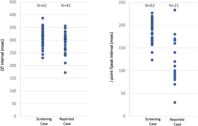
そこで我々は,既報の有症状SQTS症例と,学校心臓検診で抽出された無症状のQT短縮者の心電図所見を比較検討した.既報のSQTSはQTcと心拍数が確認できた症例42例を対象とした.学校心臓検診群では,自動診断のBazett補正またはFridericia補正がQTc<360 msとなった小1と中1の62例を対象とした.さらにJpoint-Tpeak時間も確認できた例はこれも検討した.またQT時間は接線法で再測定した値を採用した.

QT時間やQTcでは両群の重なりが多く(Fig. 7, 8),鑑別の指標には適さなかったが,Jpoint–Tpeak<120 mは検診群になく,この指標の特異度は高いと考えられた(Fig. 8).しかしSQTSではTpeakが遅い例もあり,感度は低い.

Pediatric Cardiology and Cardiac Surgery 35(1): 9-17 (2019)

Fig. 7 Distribution of corrected QT intervals between the study groups

The group of “Screening Case” comprised 1st and 7th grade children aged 6 and 12 years, respectively, who participated in a school screening program for heart disease. QTcF and QTcB indicate the QT interval corrected by Fridericia’s and Bazett’s formulas, respectively.

Pediatric Cardiology and Cardiac Surgery 35(1): 9-17 (2019)

Fig. 8 Distribution of the QT interval and J point-T peak interval between the study groups

今後の課題

SQTSの臨床像の解明は不十分であり,診断基準を提示したExpert consensus statementでもその基準に議論の余地があると述べている.成人ではQT短縮者の予後は良好と報告されているが,SQTSの心イベントは10~20歳代が多い.無症状例を含む小児SQTSの中長期予後の解明が必須である.学校心臓検診で抽出されたSQTSの予後を知ることが,学校心臓検診での抽出基準やSQTSの診断基準の適正の評価に役立つ.

また動物においては,カンガルーのQT間隔が短いことが知られ,全身麻酔時に高率に心室細動が誘発された報告がある38).人間のQT短縮でも,特殊な状況でのリスクについては検討の意義があるかもしれない.さらに心房細動既往のQT間隔が短いとの報告がある39).QT短縮者の将来の心房細動リスクも検討すべきかもしれない.

謝辞Acknowledgments

症例情報を提供してくださった以下の先生方に深謝申し上げます.

埼玉医科大学国際医療センター 住友直方 先生

大阪市立総合医療センター 江原英治 先生

滋賀医科大学 堀江稔 先生, 大野聖子 先生

埼玉県立小児医療センター 星野健司 先生

沖縄県立南部医療センター 高橋一浩 先生

独立行政法人国立病院機構 鹿児島医療センター 吉永正夫 先生

宮崎大学 高木純一 先生

千葉県循環器病センター 立野 滋 先生

利益相反

本論文について,開示すべき利益相反(COI)はない.

引用文献References

1) Priori SG, Wilde AA, Horie M, et al: HRS/EHRA/APHRS expert consensus statement on the diagnosis on the diagnosis and management of patients with inherited primary arrhythmia syndromes. Heart Rhythm 2013; 10: 1932–1963

2) Priori SG, Blomstom-Lundqvist C, Mazzanti A, et al: 2015 ESC Guidelines for the management of patients with ventricular arrhythmias and the prevention of sudden cardiac Death: The task force for management of patients with ventricular arrhythmia and the prevention of sudden cardiac death of the European society of cardiology. Eur Heart J 2015; 36: 2793–2867

3) Gussak I, Brugada P, Brugada J, et al: Idiopathic short QT interval: A new clinical syndrome? Cardiology 2000; 94: 99–102

4) Templin C, Ghadri JR, Rougier JS, et al: Identification of a novel loss-of-function calcium channel gene mutation in short QT syndrome (SQTS6). Eur Heart J 2011; 32: 1077–1088

5) Antzelevitch C, Oliva A: Amplification of spatial dispersion of repolarization underlies sudden cardiac death associated with catecholaminergic polymorphic VT, long QT, short QT and Brugada syndromes. J Intern Med 2006; 259: 48–58

6) Gollob MH, Redpath CJ, Roberts JD: The short QT syndrome: Proposed diagnostic criteria. J Am Coll Cardiol 2011; 57: 802–812

7) Patel C, Yan GX, Antzelevitch C: Short QT syndrome: From bench to bedside. Circ Arrhythm Electrophysiol 2010; 3: 401–408

8) Providência R, Karim N, Srinivasan N, et al: Impact of QTc formulae in the prevalence of short corrected QT interval and impact on probability and diagnosis of short QT syndrome. Heart 2018; 104: 502–508

9) Giustetto C, Scrocco C, Schimpf R, et al: Usefulness of exercise test in the diagnosis of short QT syndrome. Europace 2015; 17: 628–634

10) Moriya M, Seto S, Yano K, et al: Two cases of short QT interval. Pacing Clin Electrophysiol 2007; 30: 1522–1526

11) Harrell DT, Ashihara T, Ishikawa T, et al: Genotype-dependent differences in age of manifestation and arrhythmia complications in short QT syndrome. Int J Cardiol 2015; 190: 393–402

12) Sun Y, Quan XQ, Fromme S, et al: A novel mutation in the KCNH2 gene associated with short QT syndrome. J Mol Cell Cardiol 2011; 50: 433–441

13) Chinushi M, Sato A, Izumi D, et al: Nifekalant enlarged the transmural activation-recovery interval difference as well as the peak-to-end interval on surface ECG in a patient with short-QT syndrome. J Cardiovasc Electrophysiol 2012; 23: 877–880

14) Moreno C, Oliveras A, de la Cruz A, et al: A new KCNQ1 mutation at the S5 segment that impairs its association with KCNE1 is responsible for short QT syndrome. Cardiovasc Res 2015; 107: 613–623

15) Maltret A, Wiener-Vacher S, Denis C, et al: Type 2 short QT syndrome and vestibular dysfunction: Mirror of the Jervell and Lange-Nielsen syndrome? Int J Cardiol 2014; 171: 291–293

16) Righi D, Silvetti MS, Drago F: Sinus bradycardia, junctional rhythm, and low-rate atrial fibrillation in short QT syndrome during 20 years of follow-up: Three faces of the same genetic problem. Cardiol Young 2016; 26: 589–592

17) Deo M, Ruan Y, Pandit SV, et al: KCNJ2 mutation in short QT syndrome 3 results in atrial fibrillation and ventricular proarrhythmia. Proc Natl Acad Sci USA 2013; 110: 4291–4296

18) Ambrosini E, Sicca F, Brignone MS, et al: Genetically induced dysfunctions of Kir2. 1 channels: Implications for short QT3 syndrome and autism-epilepsy phenotype. Hum Mol Genet 2014; 23: 4875–4886

19) Bun SS, Maury P, Giustetto C, et al: Electrical storm in short-QT syndrome successfully treated with isoproterenol. J Cardiovasc Electrophysiol 2012; 23: 1028–1030

20) Ergül Y, Ozyilmaz I, Onan SH, et al: Short QT syndrome in a 14-year-old patient: The first pediatric case from Turkey. Anatol J Cardiol 2015; 15: 590–591

21) Mondoly P, Cardin C, Rollin A, et al: Use of a subcutaneous ICD in a patient with short QT syndrome. Clin Case Rep 2015; 4: 35–38

22) Pavao ML, Ono VC, Arfelli E, et al: Sudden cardiac death and short QT syndrome. Arq Bras Cardiol 2014; 103: e37–e40

23) Antzelevitch C, Pollevick GD, Cordeiro JM, et al: Loss-of-function mutations in the cardiac calcium channel underlie a new clinical entity characterized by ST-segment elevation, short QT intervals, and sudden cardiac death. Circulation 2007; 115: 442–449

24) Hong K, Piper DR, Diaz-Valdecantos A, et al: De novo KCNQ1 mutation responsible for atrial fibrillation and short QT syndrome in utero. Cardiovasc Res 2005; 68: 433–440

25) Suzuki H, Hoshina S, Ozawa J, et al: Short QT syndrome in a boy diagnosed on screening for heart disease. Pediatr Int 2014; 56: 774–776

26) Rollin A, Gandjbakhch E, Giustetto C, et al: Shortening of the short refractory periods in short QT syndrome. J Am Heart Assoc 2017; 6: e005684

27) Giustetto C, Schimpf R, Mazzanti A, et al: Long-term follow-up of patients with short QT syndrome. J Am Coll Cardiol 2011; 58: 587–595

28) Anttonen O, Junttila MJ, Rissanen H, et al: Prevalence and prognostic significance of short QT interval in a middle-age Finnish population. Circulation 2007; 116: 714–720

29) Gallagher MM, Magliano G, Yap YG, et al: Distribution and prognostic significance of QT intervals in the lowest half centile in 12,012 apparently healthy persons. Am J Cardiol 2006; 98: 933–935

30) Kobza R, Roos M, Niggli B, et al: Prevalence of long and short QT in a young population of 41,767 predominantly male Swiss conscripts. Heart Rhythm 2009; 6: 652–657

31) Funada A, Hayashi K, Ino H, et al: Assessment of QT intervals and prevalence of short QT syndrome in Japan. Clin Cardiol 2008; 31: 270–274

32) Miyamoto A, Hayashi H, Yoshino T, et al: Clinical and electrocardiographic characteristics of patients with short QT interval in a large hospital-based population. Heart Rhythm 2012; 9: 66–74

33) Dhutia H, Malhotra A, Parpia S, et al: The prevalence and significance of a short QT interval in 18,825 low-risk individuals including athletes. Br J Sports Med 2016; 50: 124–129

34) Guerrier K, Kwiatkowski D, Czosek RJ, et al: Short QT interval prevalence and clinical outcomes in a pediatric population. Circ Arrhythm Electrophysiol 2015; 8: 1460–1464

35) Anttonen O, Junttila J, Maury P, et al: Differences in twelve-lead electrocardiogram between symptomatic and asymptomatic subjects with short QT interval. Heart Rhythm 2009; 6: 267–271

36) Watanabe H, Makiyama T, Koyama T, et al: High prevalence of early repolarization in short QT syndrome. Heart Rhythm 2010; 7: 647–652

37) Tülümen E, Giustetto C, Wolpert C, et al: PQ segment depression in patients with short QT syndrome: A novel marker for diagnosing short QT syndrome? Heart Rhythm 2014; 11: 1024–1030

38) O’Rourke MF, Avolio AP, Nichols WW: The kangaroo as a model for the study of hypertrophic cardiomyopathy in man. Cardiovasc Res 1986; 20: 398–402

39) Poglajen G, Fister M, Radovancevic B, et al: Short QT interval and atrial fibrillation in patients without structural heart disease. J Am Coll Cardiol 2006; 47: 1905–1907

This page was created on 2019-02-22T11:10:50.406+09:00
This page was last modified on 2019-03-15T14:35:01.000+09:00


このサイトは(株)国際文献社によって運用されています。