Online ISSN: 2187-2988 Print ISSN: 0911-1794
特定非営利活動法人日本小児循環器学会 Japanese Society of Pediatric Cardiology and Cardiac Surgery
Pediatric Cardiology and Cardiac Surgery 31(3): 126-132 (2015)
doi:10.9794/jspccs.31.126

原著Original

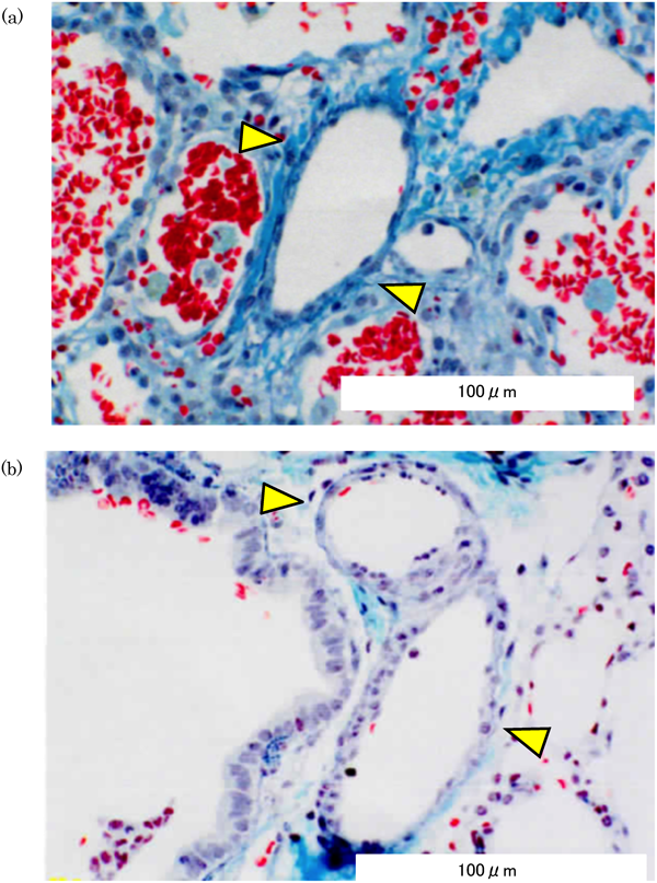
肺生検組織所見,臨床経過から考える18トリソミーの管理Management of Trisomy 18 Based on the Clinicopathology of Pulmonary Arteriopathy and Clinical Course

1土谷総合病院小児科Department of Pediatrics, Tsuchiya General Hospital ◇ 〒730-8655 広島市中区中島町3番30号3-30 Nakajima-cho, Naka-ku, Hiroshima-shi, Hiroshima 730-8655, Japan

2土谷総合病院心臓血管外科Department of Cardiovascular Surgery, Tsuchiya General Hospital ◇ 〒730-8655 広島市中区中島町3番30号3-30 Nakajima-cho, Naka-ku, Hiroshima-shi, Hiroshima 730-8655, Japan

受付日:2014年10月23日Received: October 23, 2014
受理日:2015年4月23日Accepted: April 23, 2015
発行日:2015年5月1日Published: May 1, 2015
HTMLPDFEPUB3

背景:18トリソミーの主な死因には心疾患に伴う肺出血や心不全,無呼吸などがある.

方法:高肺血流性心疾患に対して心臓手術を行った18トリソミー19人を対象に,予後を再評価し管理の参考にする目的で肺生検組織所見,経過について後方視的に検討した.

結果:日齢40.8±13.0日に肺動脈絞扼術(PAB)と肺生検を施行.肺小動脈形成不全(MD)4例(21.1%)・低形成(HPA)7例(36.8%),肺胞低形成(AH)12例(63.2%)は過去の18トリソミー以外での報告より多かった.痙攣11例.抜管した17例中閉塞性無呼吸14例.死亡6例(突然死1例,痙攣重積2例,心臓関連死(CRD)3例).PAB施行日齢とCRD, AHと術後挿管期間との間に相関を認めた.

結論:18トリソミーではMDやHPA, AHが多く,心臓関連以外にも生命予後に関わるリスクが多い.心臓手術が長期予後改善に結びつくかは不明だが,積極的治療希望の場合は,長期の人工呼吸管理を避け,生後早期に高肺血流に対するPABを行うべきである.

Background: The major causes of death in patients with trisomy 18 are heart failure, pulmonary hemorrhage because of congenital heart disease (CHD), and sudden death because of apnea.

Methods: Pulmonary artery banding (PAB) and open lung biopsy for pulmonary arterial hypertension were performed in 19 patients with trisomy 18 and CHD. The pathological findings in the small pulmonary arteries and the patients’ clinical course were analyzed.

Results: The mean age of the patients at the time of open lung biopsy was 40.8±13.0 days. Four patients (21.1%) had medial defects of the small pulmonary arteries and seven patients (36.8%) had hypoplasia of the small pulmonary arteries. Twelve patients (63.2%) had alveolar hypoplasia. The prevalence of these defects was higher in patients with trisomy 18 than in previously reported patients without trisomy 18. Eleven patients (57.8%) had associated convulsions. Overall, 14 (82.4%) of 17 patients who had been weaned from ventilatory support had obstructive apnea. Six patients died, one from sudden death, two from status epilepticus, and three from cardiac-related death (CRD). The correlation between the age at the time of PAB and CRD was significant (p<0.05), as was the correlation between the duration of artificial respiration and the grade of alveolar hypoplasia (p<0.01).

Conclusions: Medial defects and hypoplasia of the small pulmonary arteries, and alveolar hypoplasia were identified in relatively large number of patients with trisomy 18. In addition, many of these patients had risk factors affecting their prognosis. Our findings suggest that if parents request surgical intervention for their children with trisomy 18, the duration of artificial respiration should be kept as short as possible and palliative surgery should be performed as early as possible.

Key words: trisomy 18; open lung biopsy; pulmonary artery hypertension; convulsion; apnea

Ⅰ. 背景

近年,自然予後が不良とされている18トリソミーにおいて,医療的介入により短期的予後が改善されたという報告が散見される1–3).我々は,18トリソミーの肺生検組織所見を検討し,高肺血流性心疾患を伴う18トリソミーでは肺高血圧の存在にも関わらず肺小動脈の中膜肥厚が軽度であり,閉塞性肺血管病変が進行しやすく4),さらに致死的経過が予測される組織像である肺小動脈形成不全が比較的多く存在している可能性について報告した5)

医療的介入により短期的予後が改善しても,18トリソミーの長期的予後は依然として不明であり現時点では手術適応は明確にはされていない.今回,18トリソミーの肺生検組織所見に加え,合併症,死因などについて後方視的に検討し,その管理について考察したので,報告する.

Ⅱ. 方法

当院では,高肺血流性心疾患に伴う肺高血圧を呈する18トリソミーの中で,家族が積極的治療を希望した症例に対して,肺動脈絞扼術(PAB)とともに肺生検組織診断を行い,その後の方針決定の参考にしている6)

2009年から2013年の5年間に当院でPABを行った18トリソミー19例(男6例,女13例,フルトリソミー18例,モザイクトリソミー1例)(Table 1)を対象に,インフォームドコンセントの後に,全例PABと同時に肺生検を行った.PABは術中モニタリングで肺体血圧比(Pp/Ps)0.33以下を目標に,酸素飽和度や体血圧が保たれるのを確認しながら絞扼した.肺生検組織は外科的に採取しやすい肺葉から約5 mm四方のものを採取し,Heath-Edwards(HE)分類(肺血管病変分類)(1度:中膜肥厚のみ,2度:内膜細胞性増殖出現,3度:内膜線維性増殖出現,4度:肺小動脈限局性拡張,内膜叢状構造出現,5度:血管腫様病変,薄壁動脈出現,6度:壊死性血管炎(1~2度:可逆的,3~6度:不可逆的))7),IPVD(index of pulmonary vascular disease)(肺血管病変に1~4点のスコアを付けて相加平均を求めたもの:IPVD=[(1×n1)+(2×n2)+(3×n3)+(4×n4)]/(n1+n2+n3+n4),1点:内膜肥厚なし,2点:内膜細胞性肥厚あり,3点:内膜線維性肥厚あり,4点:中膜破壊あり(IPVD2.2以上は手術不可能))6,8),根治術後臨床経過区分(A: 手術後全く問題ない,B: 手術死,病院死はなく遠隔死もないが,肺高血圧が残存する,C: 手術死,病院死はないが,遠隔死の可能性が少しはある,D: 手術死はないが,病院死か遠隔死の可能性が高い,E: 手術死か病院死(A,B,C:手術適応あり,D,E:適応なし))9)を診断し,さらに肺小動脈形成不全(肺小動脈中膜が存在していない),肺小動脈低形成(肺小動脈径が並走する気管支径の60%未満),肺胞低形成(1:正常,2:肺胞壁弾性線維の軽度の欠損,3:約半数に弾性線維が欠損,4:弾性線維がほとんどない)10)についても検討した.今回,予後を再評価し管理の参考にする目的で,肺生検組織所見に加え,臨床経過として,合併症,術後挿管期間,閉塞性無呼吸,予後などについて後方視的検討を行った.

統計学的解析は,統計解析ソフトSPSS(SPSS statistics Ver. 21)を用いた.

Ⅲ. 結果(Table 1

平均在胎週数38.1±2.6週(33~41週),出生時体重1886±428 g(1,062~2,936 g).基礎心疾患は心室中隔欠損(VSD),心房中隔欠損(ASD),動脈管開存(PDA),大動脈縮窄(CoA)などであった.心エコーでの肺体血流比は1.4~6.8であり,全例肺高血圧を合併していた.平均日齢40.8±13.0日(26~69日)にPABを施行し,Pp/Psは0.90±0.16から0.42±0.16まで低下した.

Table 1 Clinical, hemodynamic and pathological findings of the patients with trisomy 18
PatientCardiac diagnosisGA (weeks)Birth weight (gram)Age at PAB (days)Qp/Qs before PABPp/Ps before PABPp/Ps after PABSite##Pathological findingsOther major complicationDuration of postoperative artificial respiration (hours)Obstructive apneaRespiratory management at hospital dischargeDischarge (months)ICR (months)Survival duration (months)
HEIPVDA–E grading*Medial defectHypoplasia of small PAAlveolar hypoplasia**
1VSD, ASD, PDA392186265.01.000.34Lul11.0A4None125+Tracheotomy138 (alive)
2#VSD, PDA382202346.01.000.38Lul11.0A1Convulsion18318 (alive)
3VSD361510365.00.700.23Rml11.0A+3None25423 (SD)
4VSD, ASD401930396.80.690.42Rml21.0A1None7+231 (alive)
5VSD, ASD411972494.21.100.30Rll01.0A2Convulsion30+3+
(15)
26 (SE)
6VSD, PDA402306342.81.080.37Rul31.7B1Forearm defect, Convulsion40+Tracheotomy78 (alive)
7CoA, VSD, ASD, PDA371746366.00.740.30Lul21.2B1None25+338 (alive)
8VSD, PDA412135387.10.570.23Lll21.1B3Convulsion25+Tracheotomy3+
(21)
52 (alive)
9VSD,ASD, PDA412246692.80.570.35Lul21.1B1Cleft lip, Convulsion12+Airway458 (alive)
10VSD, PDA381722302.70.920.67Rml31.4C3None312+3+
(9)
18 (alive)
11VSD, ASD, PDA412936344.70.930.47Rul11.0C+4Omphalocele, Convulsion312+NIPPV423 (alive)
12VSD, PDA391602472.81.000.35Rll11.0C++1CEA, Convulsion(2808)$ (CEA)+NIPPV1011 (alive)
13VSD, ASD, PDA341552201.41.000.52Llln.c.1.0D++4RDS, emphysema, ConvulsionProlonged intubation(in hospital)7 (alive)
14VSD371902292.90.860.27Lll2n.c.D+4Convulsion44+24 (SE)
15VSD, PDA331178502.70.890.30Rml21.1D+2Hepatoblastoma(4944)$$ (tracheotomy)+Tracheotomy1116 (alive)
16VSD381758622.31.000.50Rlln.c.n.c.D++1HPS14327 (CRD)
17VSD, PDA371788362.21.000.66Lll32.0E+4Convulsion312+NIPPV525 (alive)
18VSD, PDA402102443.51.000.70Lul31.6E3CDH, Convulsion12+NIPPV7+
(14)
14 (CRD)
19CoA, VSD, ASD, PDA331062624.51.000.68Lll31.5E2CEA, Imperforate anusProlonged intubation(in hospital)7 (CRD)
VSD: ventricular septal defect, ASD: atrial septal defect, PDA: patent ductus arteriosus, CoA: coarctation of aorta, GA: gestational age, Qp/Qs: ratio of pulmonary to systemic flow, PAB: pulmonary artery banding, Pp/Ps: ratio of pulmonary to systemic pressure, HE: Heath Edwards classification, n.c.: not classifiable, IPVD: index of pulmonary vascular diesase, PA: pulmonary artery, CEA: congenital esophageal atresia, RDS: respiratory distress syndrome, HPS: hypertrophic pyloric stenosis, CDH: congenital diaphragmatic hernia, NIPPV: non-invasive positive pressure ventilation, ICR: intracardiac repair, SD: sudden death, SE: death associated with status epileptics, CRD: cardiac-related death. #: mosaic trisomy 18, Site. ##: the site of pulmonary biopsy (R: right, L: left, ul: upper lobe, ml: mid lobe, ll: lower lobe), A–E grading*: postoperative predictable prognosis (A: absolutely no postoperative problems, B: no operative or in-hospital death, but pulmonary hypertension remains, C: no operative or in-hospital death, but late death is possible, D: no operative or in-hospital death, but late death is likely, E: operative or in-hospital death), Alveolar hypoplasia** (grade 1: the alveolar structure is sufficiently reinforced by entrance rings containing the normal quantity of elastic fibres, grade 2: mild deficiency of elastic fibres, grade 3: the rings have only half the normal amount of elastic fibres, grade 4: the entrance rings have only a small number of elastic fibres), $, $$: the duration of postoperative artificial respiration was long because of CEA, stenosis of the trachea, respectively.

肺生検組織所見では,HE分類は0度1例,1度5例,2度6例,3度5例で肺小動脈形成不全のために分類不能だったものが2例あった.IPVDは1.0~2.0で,根治術後臨床経過区分はA:5例,B:4例,C:3例,D:4例,E:3例であった.肺小動脈形成不全は4例(21.1%)に認め(Figure 1(a), 1(b)),肺小動脈低形成は7例(36.8%)に認めた(Figure 2).一方,肺胞低形成は1度7例,2度3例,3度4例,4度5例であり,2度以上は12例(63.2%)であった(Figure 3).

Pediatric Cardiology and Cardiac Surgery 31(3): 126-132 (2015)

Fig. 1 Medial defects of the small pulmonary arteries visualized by Elastica Goldner staining in (a) Patient 12 and in (b) Patient 14

Pediatric Cardiology and Cardiac Surgery 31(3): 126-132 (2015)

Fig. 2 Hypoplasia of the small pulmonary arteries visualized by Elastica Goldner staining in Patient 11

Pediatric Cardiology and Cardiac Surgery 31(3): 126-132 (2015)

Fig. 3 Grade 4 alveolar hypoplasia visualized by Elastica Goldner staining in Patient 1

5例(26.3%)に消化管奇形を合併しており(臍帯ヘルニア,食道閉鎖,肥厚性幽門狭窄,先天性横隔膜ヘルニア,食道閉鎖+低位鎖肛),全例他院で外科的介入が行われた後に紹介になっていたため,PAB施行日齢が遅い傾向があった(日齢34~62).11例(57.9%)に痙攣を認め,そのうち2例は痙攣重積に続発した無呼吸で死亡した.術後挿管期間は7~4,944時間であった(2例は未抜管).このうち,症例12,19は食道閉鎖根治術待機のため,症例15は気道狭窄のため気管切開施行まで計画的に長期挿管となっており,症例13,19は肺気腫,肺高血圧合併などのため換気条件が下げられない状態であった.人工呼吸器から離脱した17例中14例(82.4%)に閉塞性無呼吸を認め,退院時に4例が気管切開,4例がNIPPV(Non-invasive positive pressure ventilation),1例がエアウェイを必要とした.抜管困難の2例を除いて平均4.4±2.8ヶ月で退院した.過去の報告では1年生存率は5~10%とされているが11,12),今回の検討では生存者の経過年数は1年生存4人,2年生存2人,3年生存2人,4年生存2人であり,Kaplan–Meier法による生存曲線では1年生存率は89.4%であった(Figure 4).亡くなった6例の死因は,突然死1例,痙攣重積2例,心臓関連死3例であった.

Pediatric Cardiology and Cardiac Surgery 31(3): 126-132 (2015)

Fig. 4 Survival curve for postoperative patients with trisomy 18 with pulmonary arterial hypertension because of congenital heart disease

統計学的解析ではPAB施行日齢と心臓関連死の間に正の相関を認め(p<0.05),肺生検組織所見では肺胞低形成と術後挿管期間の間に正の相関を認めていた(p<0.01)(Table 2).

Table 2 Correlation between the pathological findings and the clinical course of trisomy 18
The clinical courseDuration of postoperative artificial respiration*Obstructive apnea**ConvulsionCardiac-related death
Age at PABn.s.#n.s.##n.s.##p<0.05##
HE***n.s.##n.s.####n.s.####n.s.####
IPVD***n.s.#n.s.##n.s.##n.s.##
A–E gradingn.s.###n.s.##n.s.##n.s.##
Medial defectn.s.##n.s.####n.s.####n.s.####
Hypoplasia of small PAn.s.##n.s.####n.s.####n.s.####
Alveolar hyoplasiap<0.01###n.s.##n.s.##n.s.##
PAB: pulmonary artery banding, HE: Heath Edwards classification, IPVD: Index of pulmonary vascular disease, A–E grading: postoperative predictable prognosis, PA: pulmonary artery, n.s.: not significant. *: Correlation analysis of the patients with trisomy 18 other than patients receiving prolonged mechanical ventilation. **: Correlation analysis of the patients with trisomy 18 other than patients that had not been weaned from mechanical ventilation. ***: Correlation analysis of the patients with trisomy 18 that could be classified by category, respectively. #: Pearson’s correlation coefficient, ##: one-way analysis of variance (ANOVA), ###: Spearman’s rank correlation coefficient, ####: Cramer’s coefficient of association.

Ⅳ. 考察

18トリソミーは3,500~8,500人に1人の頻度で出生する染色体異常症であり,重度成長発達遅滞,先天性心疾患などを合併し,1年生存率は5~10%とされている11,12).心疾患に関する本邦での報告では,18トリソミーの94%は先天性心疾患を合併し,うち52%が肺高血圧を呈していた13).25%に心臓手術が行われ,ほとんどは姑息術であり,手術施行例の19%に心内修復術が行われていた13).主な死因として,Koshoらは先天性心疾患・心不全,肺高血圧を背景に,突発的な呼吸心停止または肺高血圧に関連したエピソードで亡くなる児が多いと報告しており14),Ceredaらは心疾患に伴う心不全や肺出血,中枢性無呼吸,呼吸不全,誤嚥,閉塞性無呼吸などを挙げている15)

高肺血流性心疾患では肺血流増加に伴う肺動脈圧の上昇に対し,肺小動脈中膜が肥厚することで,内膜増殖性病変の進行を阻止するが16,17),18トリソミーでは肺小動脈の中膜肥厚が軽度であり,肺血管閉塞性病変が早期に進行しやすく4),この結果肺高血圧の早期増悪を来し死に至る可能性がある18,19).今回の検討では,肺小動脈形成不全を21.1%,肺小動脈低形成を36.8%に認めた.Yamakiは先天性心疾患を合併した18,13トリソミー以外の児では肺小動脈形成不全を385例中2例(0.5%)に認め,VSDもしくはASDもしくはPDAを合併した21トリソミーでは肺小動脈低形成を94例中3例(3.2%)に認めたと報告している20).年齢,体格,肺動脈圧などの因子にばらつきがあり,今回のデータと単純に比較できないが,18トリソミーに肺小動脈形成不全,肺小動脈低形成を認める頻度は高いと考えられる(Table 3).Kanekoらは薬物療法と手術を組み合わせることで,生命予後の改善が期待できる可能性があり,心臓手術が心臓関連死の予防において有用であることを示唆している1,2).今回の検討でもPAB施行日齢と心臓関連死との間に正の相関を認めており,より早期にPABを施行することで閉塞性肺血管病変の進行が予防でき,少なくとも心臓関連死のリスクを減少することが期待できる.しかし,心臓手術が18トリソミーの長期的な生命予後を改善するか否かは依然として不明である13,21).肺小動脈形成不全,肺小動脈低形成の存在は予後に負に働くことが推察されることから,18トリソミーでは早期手術により閉塞性肺血管病変の進行を予防しても,長期的な生命予後の改善が期待できない症例が比較的多く存在していると考えられる.

Table 3 Pathological findings: Comparison of trisomy 18 with other groups
Patient with PAH associated with CHDnMedial defectHypoplasia of small pulmonary arteryAlveolar hyoplasia
18 trisomy194 (21.1%)7 (36.8%)12 (63.2%)
No chromosome abnormalities20)3852 (0.5%)*
21 trisomy20)943 (3.2%)*
21 trisomy10)2824 (85.7%)**
No chromosome abnormalities10)244 (16.7%)*
PAH: pulmonary artery hypertension, CHD: congenital heart disease. *: p<0.01 vs. trisomy 18, **: no significant different vs. trisomy 18.

一方,今回の検討では肺胞低形成を63.2%に認めた.Yamakiらは先天性心疾患を合併した21トリソミーでは肺胞低形成を28例中24例(85.7%)に認め,先天性心疾患を合併した21トリソミー以外の児では24例中4例(16.7%)に認めたと報告しており10),18トリソミーでは,染色体異常のない児と比較して肺胞低形成の頻度が高いと考えられる(Table 3).また,肺胞低形成と術後挿管期間との間に高い正の相関を認めている(Table 2).肺胞壁中の弾性線維は呼吸における肺の弾性収縮に関わっており,18トリソミーでは術後の呼吸不全や肺気腫などのリスクが高いと考えられる.Yamakiらは21トリソミーでの検討で同様の所見を得ている10).呼吸器症状や術後管理に与える影響について18トリソミーと21トリソミーでの違いは,臨床的背景などの違いもあり,一概に論じることはできない.今後の症例の蓄積によって両者間の違いを検討していくことが望ましい.

消化管奇形を合併した5例は,外科的介入後のためPAB施行日齢が比較的遅く,根治術後臨床経過区分はそれぞれC~Eに分類され,5例中3例は心臓関連で死亡している.Koshoらは食道閉鎖の存在自体は,根治術を行うことができれば必ずしも絶対的生命予後不良因子にはならないとしている14).より早期にPABを施行できるように努めることで,短期的予後の改善が期待できる.

痙攣は11例(57.9%)に認めた.うち2例は抗痙攣剤投与中にも関わらず,痙攣重積で死亡しており,18トリソミーの予後に関わる重大な合併症と言える.一方,てんかん性無呼吸は発作時脳波で無呼吸に先行する異常波の出現で診断されるが,乳児期以降に発症することは稀とされている22).Kumadaらは18トリソミーにおけるてんかん性無呼吸の存在について報告しており,亡くなった2例とも痙攣後に自宅で呼吸停止したのであるが,てんかん性無呼吸であった可能性は否定できない.中枢性無呼吸の治療は痙攣を誘発するリスクがあり,てんかん性無呼吸と中枢性無呼吸との鑑別は重要である.軽微な所見であっても早期から痙攣に対する積極的な検査と治療を行う必要がある23,24)

閉塞性無呼吸は82.4%に認めた.Koshoらは47例中24例(51%)に無呼吸を認め,12例に用手的刺激,3例に人工呼吸を要したとしている24).近年,気管切開を含めた治療を行う施設が増えてきており25),適切な呼吸管理を行うことで,少なくとも閉塞性無呼吸に伴う死亡は回避できる可能性はある.

18トリソミーでは早期に手術を行うことで心臓関連死を予防できることが期待できるが,一方で肺小動脈形成不全,肺小動脈低形成,肺胞低形成などの予後に負に働くことが推察される肺組織所見を有する児が比較的多かった.また,18トリソミーは手術危険率が高く2,21),その特徴的肺組織所見からは体外循環のリスクもかなり高いことが予想される.生後早期であれば姑息術であるPABを選択するべきと考えた.さらに生後早期からの外科的介入によって閉塞性肺血管病変の進行が予防できても,痙攣や閉塞性無呼吸といった生命予後に関わる心臓関連以外のリスクも多く,依然として18トリソミーの長期的予後は不明であると言わざるをえない.18トリソミーを持つ親は,出生直後から生命予後の厳しさや新生児期の蘇生,手術などの現実的な決定事項に直面し,その後生存している場合には重度の発達遅延と向き合わなければならない.18トリソミーの手術適応を含めた治療方針の決定には,両親の感情や思考に対して敬意を持って,両親と共に児の最善の利益を追求する姿勢で検討することが大切である.

Ⅴ. 結語

高肺血流性心疾患を伴う18トリソミーの肺生検組織所見では,肺小動脈形成不全や肺小動脈低形成,肺胞低形成が比較的多く認められた.また,18トリソミーには痙攣や無呼吸などの生命予後に関わる心臓関連以外のリスクも多く,依然として長期的予後は不明である.高肺血流性心疾患を伴う18トリソミーの場合,心臓手術が必ずしも長期的予後改善に結びつくとは限らないが,積極的治療を希望される場合には,心臓関連死を予防し,退院して在宅医療へ移行するためにも,長期の人工呼吸管理を避け,生後早期に心臓姑息術であるPABを行うべきである.

本研究の限界

本研究は積極的治療を希望されて,実際に手術を行った18トリソミーを対象とした研究であり,手術を受けなかった児との比較検討が行えていない.今回の知見を18トリソミー共通のものとするには,今後のさらなる症例の集積とともに,手術を受けなかった児との臨床的背景も含めた比較検討が必要である.

謝辞Acknowledgments

本稿を作成するにあたり,肺生検組織診断をして頂いた日本肺血管研究所の八巻重雄先生,統計学的解析を監修して頂いた広島赤十字・原爆病院内分泌・代謝内科の中西修平先生に深く感謝申し上げます.

引用文献References

1) Kaneko Y, Kobayashi J, Yamamoto Y, et al: Intensive cardiac management in patients with trisomy 13 or trisomy 18. Am J Med Genet A 2008; 146A: 1372–1380

2) Kaneko Y, Kobayashi J, Achiwa I, et al: Cardiac surgery in patients with trisomy 18. Pediatr Cardiol 2009; 30: 729–734

3) Kobayashi J, Kaneko Y, Yamamoto Y, et al: Radical surgery for a ventricular septal defect associated with trisomy 18. Gen Thorac Cardiovasc Surg 2010; 58: 223–227

4) 田原昌博,本田 茜,下薗彩子,ほか:高肺血流性心疾患を伴う18トリソミーの肺生検組織所見.日児誌2013; 117: 1260–1266

5) Tahara M, Shimozono S, Nitta T, et al: Medial defects of the small pulmonary arteries in fatal pulmonary hypertension in infants with trisomy 13 and trisomy 18. Am J Med Genet A 2014; 164A: 319–323

6) Yamaki S, Mohri H, Haneda K, et al: Indication for surgery based on lung biopsy in cases of ventricular septal defect and/or patent ductus arteriosus with severe pulmonary hypertension. Chest 1989; 96: 31–39

7) Heath D, Edwards JE: The pathology of hypertensive pulmonary vascular disease: A description of six grades of structural changes in the pulmonary arteries with special reference to congenital cardiac septal defects. Circularion 1958; 18: 533–547

8) Yamaki S, Tezuka F: Quantitative analysis of pulmonary vascular disease in complete transposition of great arteries. Circulation 1976; 54: 805–809

9) Yamaki S: Lung biopsy diagnosis of pulmonary vascular disease. Tokyo, Japan: Medical Review, 2000, 89–92

10) Yamaki S, Horiuchi T, Takahashi T: Pulmonary changes in congenital heart disease with Down’s syndrome: Their significance as a cause of postoperative respiratory failure. Thorax 1985; 40: 380–386

11) Edwards JUH, Harnden DG, Cameron AH, et al: A new trisomic syndrome. Lancet 1960; 1: 787–789

12) Rasmussen SA, Wong LY, Yang Q, et al: Population-based analysis of mortality in trisomy 13 and trisomy 18. Pediatrics 2003; 111: 777–784

13) Maeda J, Yamagishi H, Furutani Y, et al: The impact of cardiac surgery in patients with trisomy 18 and trisomy 13 in Japan. Am J Med Genet A 2011; 155A: 2641–2646

14) Kosho T, Nakamura T, Kawame H, et al: Neonatal management of trisomy 18: Clinical details of 24 patients receiving intensive treatment. Am J Med Genet 2006; 140A: 937–944

15) Cereda A, Carey JC: The trisomy 18 syndrome. Orphanet J Rare Dis 2012; 7: 81

16) Yamaki S, Wagenvoort CA: Plexogenic pulmonary arteriopathy: Significance of medial thickness with respect to advanced pulmonary vascular lesions. Am J Pathol 1981; 105: 70–75

17) Yamaki S, Abe A, Tabayashi K, et al: Inoperable pulmonary vascular disease in infants with congenital heart disease. Ann Thorac Surg 1998; 66: 1565–1570

18) Van Praagh S, Truman T, Firpo A, et al: Cardiac malformations in trisomy-18: A study of 41 postmortem cases. J Am Coll Cardiol 1989; 13: 1586–1597

19) Musewe NN, Alexander DJ, Teshima I, et al: Echocardiographic evaluation of the spectrum of cardiac anomalies associated with trisomy 13 and trisomy 18. Pediatr Cardiol 1990; 15: 673–677

20) Yamaki S: Pulmonary vascular disease associated with pulmonary hypertension in 445 patients: Diagnosis from lung biopsy and autopsy. Gen Thorac Cardiovasc Surg 2013; 61: 24–31

21) Yamagishi H: Cardiovascular surgery for congenital heart disease associated with trisomy 18. Gen Thorac Cardiovasc Surg 2010; 58: 217–219

22) Ramelli GP, Donati F, Bianchetti M, et al: Apnoeic attacks as an isolated manifestation of epileptic seizures in infants. Eur J Paediatr Neurol 1998; 2: 187–191

23) Kumada T, Nishii R, Higashi T, et al: Epileptic apnea in a trisomy 18 infant. Pediatr Neurol 2010; 42: 61–64

24) Kosho T, Kuniba H, Tanikawa Y, et al: Natural history and parental experience of children with trisomy 18 based on a questionnaire given to a Japanese trisomy 18 parental support group. Am J Med Genet A 2013; 161A: 1531–1542

25) Nelson KE, Hexem KR, Feudtner C: Inpatient hospital care of children with trisomy 13 and trisomy 18 in the United States. Pediatrics 2012; 129: 869–876

This page was created on 2015-04-28T14:44:26.653+09:00
This page was last modified on 2015-05-25T09:10:19.327+09:00


このサイトは(株)国際文献社によって運用されています。